Human TSLPR Antibody
R&D Systems, part of Bio-Techne | Catalog # AF981


Key Product Details
Validated by
Species Reactivity
Validated:
Cited:
Applications
Validated:
Cited:
Label
Antibody Source
Product Specifications
Immunogen
Val30-Phe232
Accession # Q9HC73
Specificity
Clonality
Host
Isotype
Endotoxin Level
Scientific Data Images for Human TSLPR Antibody
Detection of Human TSLP R by Western Blot.
Western blot shows lysates of NS0 mouse myeloma cell line either mock transfected or transfected with human TSLP R. PVDF membrane was probed with 0.2 µg/mL of Goat Anti-Human TSLP R Antigen Affinity-purified Polyclonal Antibody (Catalog # AF981) followed by HRP-conjugated Anti-Goat IgG Secondary Antibody (Catalog # HAF017). Specific bands were detected for TSLP R at approximately 45-60 kDa (as indicated). This experiment was conducted under reducing conditions and using Immunoblot Buffer Group 1.Detection of Human TSLP R by Western Blot.
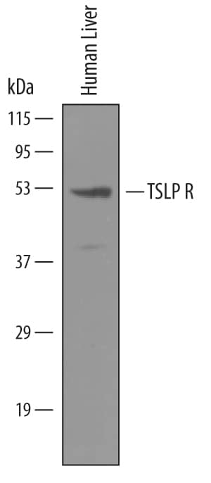
Western blot shows lysates of human liver tissue. PVDF membrane was probed with 1 µg/mL of Goat Anti-Human TSLP R Antigen Affinity-purified Polyclonal Antibody (Catalog # AF981) followed by HRP-conjugated Anti-Goat IgG Secondary Antibody (Catalog # HAF019). A specific band was detected for TSLP R at approximately 50 kDa (as indicated). This experiment was conducted under reducing conditions and using Immunoblot Buffer Group 8.TSLP R in Human Lymph Node.
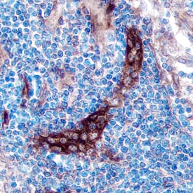
TSLP R was detected in immersion fixed paraffin-embedded sections of human lymph node using Goat Anti-Human TSLP R Antigen Affinity-purified Polyclonal Antibody (Catalog # AF981) at 5 µg/mL overnight at 4 °C. Tissue was stained using the Anti-Goat HRP-DAB Cell & Tissue Staining Kit (brown; Catalog # CTS008) and counter-stained with hematoxylin (blue). Specific staining was localized to the plasma membrane. View our protocol for Chromogenic IHC Staining of Paraffin-embedded Tissue Sections.Applications for Human TSLPR Antibody
Immunocytochemistry
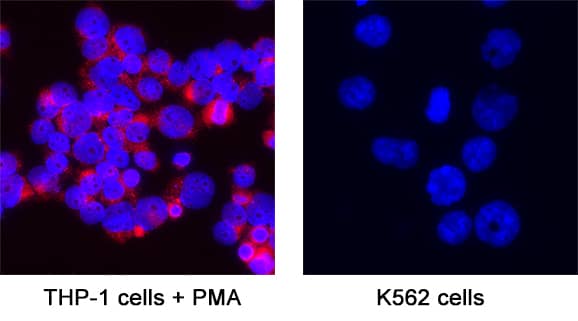
Sample: Immersion fixed THP‑1 human acute monocytic leukemia cell line treated with PMA
Immunohistochemistry
Sample: Immersion fixed paraffin-embedded sections of human lymph node
Western Blot
Sample: Human liver tissue, NS0 mouse myeloma cell line either mock transfected or transfected with human TSLP R
Neutralization
Reviewed Applications
Read 2 reviews rated 4 using AF981 in the following applications:
Formulation, Preparation, and Storage
Purification
Reconstitution
Formulation
*Small pack size (-SP) is supplied either lyophilized or as a 0.2 µm filtered solution in PBS.
Shipping
Stability & Storage
Background: TSLPR
TSLP R, also named Delta (1) and CRLM-2 (2) (cytokine receptor-like module-2), was originally cloned as a novel type 1 cytokine receptor with similarity to the common gamma chain. It was subsequently identified to be a subunit of the cellular receptor for the IL-7-like cytokine TSLP and termed TSLP R (3). The human TSLP R cDNA encodes a 371 amino acid (aa) residue type 1 membrane protein with a 22 aa residue signal peptide, a 210 aa residue extracellular domain, a 20 aa residue transmembrane domain, and a 119 aa residue cytoplasmic domain (4, 5). The extracellular region contains two fibronectin type III-like domains and a WSXWS‑like motif. The cytoplasmic domain contains a membrane-proximal box 1 motif that is known to be important for association with JAKs (4). Human TSLP R displays 39% identity to mouse TSLP R and 24% identity to the common gamma receptor (4). An alternatively spliced mRNA variant encoding a soluble TSLP R has also been reported in mouse (2). TSLP R expression is ubiquitous in the immune and hematopoietic cells, but is up-regulated in Th2-skewed cells. Cells expressing TSLP R alone bind TSLP with low affinity. Co-expression of TSLP R and IL-7 R alpha is required for high-affinity TSLP binding and signal transduction (3-6). The TSLP R and IL-7 R alpha are coexpressed primarily on monocytes and dendritic cells and at lower levels in lymphoid cells. TSLP has been shown to induce the release of T cell‑attracting chemokines from monocytes and enhance the maturation of CD11c+ dendritic cells (5).
References
- Fujio, K. et al. (2000) Blood 95:2204.
- Hiroyama, T. et al. (2000) Biochem. Biophys. Res. Commun. 272:224.
- Park, L.S. et al. (2000) J. Exp. Med. 192:659.
- Tonozuka, Y. et al. (2001) Cytogenet. Cell Genet. 93:23.
- Reche, P.A. et al. (2001) J. Immunol. 167:336.
- Pandey, A. et al. (2000) Nat. Immunol. 1:59.
Long Name
Alternate Names
Gene Symbol
UniProt
Additional TSLPR Products
Product Documents for Human TSLPR Antibody
Product Specific Notices for Human TSLPR Antibody
For research use only


