Human VAP-1/AOC3 Antibody
R&D Systems, part of Bio-Techne | Catalog # AF3957

Key Product Details
Species Reactivity
Applications
Label
Antibody Source
Product Specifications
Immunogen
Gly27-Asn763
Accession # Q16853
Specificity
Clonality
Host
Isotype
Scientific Data Images for Human VAP-1/AOC3 Antibody
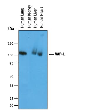
Detection of Human VAP-1/AOC3 by Western Blot.
Western blot shows lysates of human lung tissue, human kidney tissue, human liver tissue, and human heart tissue. PVDF membrane was probed with 1 µg/mL of Goat Anti-Human VAP-1/AOC3 Antigen Affinity-purified Polyclonal Antibody (Catalog # AF3957) followed by HRP-conjugated Anti-Goat IgG Secondary Antibody (Catalog # HAF019). A specific band was detected for VAP-1/AOC3 at approximately 95-100 kDa (as indicated). This experiment was conducted under reducing conditions and using Immunoblot Buffer Group 1.Detection of Human VAP‑1/AOC3 by Simple WesternTM.
Simple Western lane view shows lysates of human lung tissue and heart tissue, loaded at 0.2 mg/mL. A specific band was detected for VAP-1/AOC3 at approximately 123 kDa (as indicated) using 10 µg/mL of Goat Anti-Human VAP-1/AOC3 Antigen Affinity-purified Polyclonal Antibody (Catalog # AF3957) followed by 1:50 dilution of HRP-conjugated Anti-Goat IgG Secondary Antibody (Catalog # HAF109). This experiment was conducted under reducing conditions and using the 12-230 kDa separation system.Applications for Human VAP-1/AOC3 Antibody
Simple Western
Sample: Human lung tissue and heart tissue
Western Blot
Sample: Human lung tissue, human liver tissue, and human heart tissue
Formulation, Preparation, and Storage
Purification
Reconstitution
Formulation
Shipping
Stability & Storage
- 12 months from date of receipt, -20 to -70 °C as supplied.
- 1 month, 2 to 8 °C under sterile conditions after reconstitution.
- 6 months, -20 to -70 °C under sterile conditions after reconstitution.
Background: VAP-1/AOC3
Vascular Adhesion Protein-1 (VAP-1) is a copper amine oxidase with a topaquinone cofactor. VAP-1 is a Type II integral membrane protein, but a soluble form of the enzyme is present in human serum, and its level increases in diabetes and some inflammatory liver diseases (1, 2). VAP-1 catalyzes the oxidative deamination of small primary amines such as methylamine, benzylamine, and aminoacetone in a reaction that produces an aldehyde, ammonia, and H2O2 (3). The enzyme is sensitive to inhibition by semicarbazide. VAP-1 expression is highest in the endothelium of lung, heart, and intestine, but low in tissues such as brain, spleen, kidney, and liver (4). VAP-1 vascular expression is regulated at sites of inflammation through its release from intracellular granules in which the protein is stored (5). The adhesive function of VAP-1 has been demonstrated in studies showing that the protein is important for the adherence of certain lymphocyte subtypes to inflamed endothelial tissues (6). VAP-1 mediated adhesion is involved in the process of leukocyte extravasation, an important feature of inflammatory responses. The role of VAP-1 amine oxidase activity in this process is not fully defined, but it appears to be carbohydrate-dependent (7). VAP-1 is considered to be a therapeutic target for diabetes, oxidative stress, and inflammatory diseases (8).
References
- Kurkijärvi, R. et al. (1998) J. Immunol. 161:1549.
- Gearing, A.J.H. and W. Newman (1993) Immunol. Today 14:506.
- Lizcano, J.M. et al. (1998) Biochem. J. 331:69.
- Smith, D.J. et al. (1998) J. Exp. Med. 188:17.
- Jaakkala K. et al. (2000) Am. J. Pathol. 157:463.
- Salmi, M. and J. Jalkanen (2001) Trends Immunol. 22:211.
- Salmi, M. and J. Jalkanen (1996) J. Exp. Med. 183:569.
- Dunkel, P. et al. (2008) Curr. Med. Chem. 15:1827.
Long Name
Alternate Names
Gene Symbol
UniProt
Additional VAP-1/AOC3 Products
Product Documents for Human VAP-1/AOC3 Antibody
Product Specific Notices for Human VAP-1/AOC3 Antibody
For research use only

