Human VEGFR1/Flt-1 Antibody
R&D Systems, part of Bio-Techne | Catalog # AF321


Key Product Details
Species Reactivity
Validated:
Human
Cited:
Human, Bovine
Applications
Validated:
Blockade of Receptor-ligand Interaction, CyTOF-ready, Flow Cytometry, Immunohistochemistry, Western Blot
Cited:
Blocking, Flow Cytometry, Functional Assay, Immunocytochemistry, Immunohistochemistry, Immunohistochemistry-Frozen, Immunohistochemistry-Paraffin, Neutralization, Proximity Ligation Assay (PLA), Western Blot
Label
Unconjugated
Antibody Source
Polyclonal Goat IgG
Product Specifications
Immunogen
S. frugiperda insect ovarian cell line Sf 21-derived recombinant human VEGFR1/Flt-1
Ser27-His687
Accession # NP_001153392
Ser27-His687
Accession # NP_001153392
Specificity
Detects human VEGFR1/Flt-1 in direct ELISAs and Western blots.
Clonality
Polyclonal
Host
Goat
Isotype
IgG
Endotoxin Level
<0.10 EU per 1 μg of the antibody by the LAL method.
Scientific Data Images for Human VEGFR1/Flt-1 Antibody
VEGFR1/Flt‑1 in Human Breast Cancer Tissue.
VEGFR1/Flt‑1 was detected in immersion fixed paraffin-embedded sections of human breast cancer tissue using 15 µg/mL Human VEGFR1/Flt‑1 Antigen Affinity-purified Polyclonal Antibody (Catalog # AF321) overnight at 4 °C. Tissue was stained (red) and counterstained with hematoxylin (blue). View our protocol for Chromogenic IHC Staining of Paraffin-embedded Tissue Sections.VEGFR1/Flt‑1 in Human Ovarian Cancer Tissue.
VEGFR1/Flt-1 was detected in immersion fixed paraffin-embedded sections of human ovarian cancer tissue using Human VEGFR1/Flt-1 Antigen Affinity-purified Polyclonal Antibody (Catalog # AF321) at 3 µg/mL overnight at 4 °C. Tissue was stained using the Anti-Goat HRP-DAB Cell & Tissue Staining Kit (brown; CTS008) and counterstained with hematoxylin (blue). View our protocol for Chromogenic IHC Staining of Paraffin-embedded Tissue Sections.Detection of Human VEGFR1/Flt-1 by Western Blot
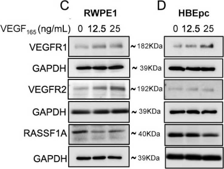
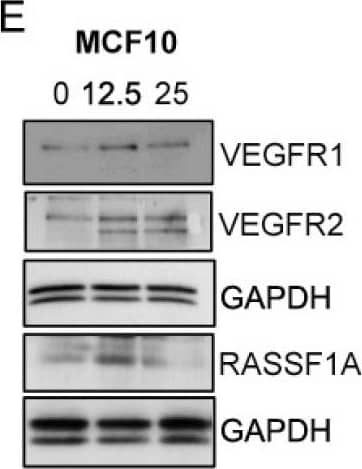
Exogenous VEGF165 suppresses RASSF1A expression in normal epithelial and endothelial cells. Metastatic colon cancer cells (mCRC, (A) T84 and (B) Colo 205) were stimulated with VEGF165 as indicated before and cell metabolism was performed by MTT assay for 6 h (black dash), 24 h (green) and 48 h (pink). Immortalized benign protate ((C) RWPE1), human bronchial epithelial cells ((D) HBEpc), immortalized human breast epithelial cells ((E) MCF-10A), endothelial cells (F, EC) were stimulated with or without VEGF165 (12.5 and 25 ng/mL) for 24 h. Cell lysates were monitored by Western blot for p-VEGFR1, p-VEGFR2, RASSF1A, and GAPDH as indicated. Immunoblot was quantified by scanning densitometry and normalized against GAPDH expression for RWPE1 (G), HBEpc (H), MCF 10A (I) and EC (lower panel of (F)). Results are from three independent experiments and statistical significance was determined using one way-ANOVA followed Bonferroni test. (* p < 0.05, ** p < 0.01, *** p < 0.001). Image collected and cropped by CiteAb from the following open publication (https://pubmed.ncbi.nlm.nih.gov/30744076), licensed under a CC-BY license. Not internally tested by R&D Systems.Applications for Human VEGFR1/Flt-1 Antibody
Application
Recommended Usage
Blockade of Receptor-ligand Interaction
In a functional ELISA, 1-4 µg/mL of this antibody will block 50% of the binding of 2 ng/mL of Recombinant Human PlGF (Catalog # 264-PG) to immobilized Recombinant Human VEGFR1/Flt-1 Fc Chimera (Catalog # 321-FL) coated at 1 µg/mL (100 µL/well). At 30 μg/mL, this antibody will block >90% of the binding.
CyTOF-ready
Ready to be labeled using established conjugation methods. No BSA or other carrier proteins that could interfere with conjugation.
Flow Cytometry
0.25 µg/106 cells
Sample: HUVEC human umbilical vein endothelial cells
Sample: HUVEC human umbilical vein endothelial cells
Immunohistochemistry
5-15 µg/mL
Sample: Immersion fixed paraffin-embedded sections of human breast cancer and ovarian cancer tissues
Sample: Immersion fixed paraffin-embedded sections of human breast cancer and ovarian cancer tissues
Western Blot
0.1 µg/mL
Sample: Recombinant Human VEGFR1/Flt‑1 Fc Chimera (Catalog # 321-FL)
Sample: Recombinant Human VEGFR1/Flt‑1 Fc Chimera (Catalog # 321-FL)
Reviewed Applications
Read 7 reviews rated 4.6 using AF321 in the following applications:
Formulation, Preparation, and Storage
Purification
Antigen Affinity-purified
Reconstitution
Reconstitute at 0.2 mg/mL in sterile PBS. For liquid material, refer to CoA for concentration.
Formulation
Lyophilized from a 0.2 μm filtered solution in PBS with Trehalose. See Certificate of Analysis for details.
*Small pack size (-SP) is supplied either lyophilized or as a 0.2 µm filtered solution in PBS.
*Small pack size (-SP) is supplied either lyophilized or as a 0.2 µm filtered solution in PBS.
Shipping
Lyophilized product is shipped at ambient temperature. Liquid small pack size (-SP) is shipped with polar packs. Upon receipt, store immediately at the temperature recommended below.
Stability & Storage
Use a manual defrost freezer and avoid repeated freeze-thaw cycles.
- 12 months from date of receipt, -20 to -70 °C as supplied.
- 1 month, 2 to 8 °C under sterile conditions after reconstitution.
- 6 months, -20 to -70 °C under sterile conditions after reconstitution.
Background: VEGFR1/Flt-1
Vascular Endothelial Growth Factor Receptor 1 (VEGFR1) is a receptor tyrosine kinase that is expressed primarily on endothelial cells and plays a role in vasculogenesis and angiogenesis. A soluble variant of VEGFR1 was also reported to bind VEGF and PlGF with high affinity and function as a potent VEGF antagonist.
Long Name
Vascular Endothelial Growth Factor Receptor 1
Alternate Names
Flt-1, FLT1, FRT, VEGF R1, VEGFR-1
Gene Symbol
FLT1
UniProt
Additional VEGFR1/Flt-1 Products
Product Documents for Human VEGFR1/Flt-1 Antibody
Product Specific Notices for Human VEGFR1/Flt-1 Antibody
For research use only
Loading...
Loading...
Loading...
Loading...
Loading...


