Human VMAT2 Antibody
R&D Systems, part of Bio-Techne | Catalog # MAB8327


Key Product Details
Species Reactivity
Applications
Label
Antibody Source
Product Specifications
Immunogen
Met1-Asp514
Accession # Q05940
Specificity
Clonality
Host
Isotype
Scientific Data Images for Human VMAT2 Antibody
Detection of Human VMAT2 by Western Blot.
Western blot shows lysates of HEK293 human embryonic kidney cell line either mock transfected or transfected with human VMAT2. PVDF membrane was probed with 1 µg/mL of Mouse Anti-Human VMAT2 Monoclonal Antibody (Catalog # MAB8327) followed by HRP-conjugated Anti-Mouse IgG Secondary Antibody (Catalog # HAF018). A specific band was detected for VMAT2 at approximately 75-100 kDa (as indicated). This experiment was conducted under reducing conditions and using Immunoblot Buffer Group 1.Detection of VMAT2 in HEK293 Human Cell Line Transfected with Human VMAT2 and eGFP by Flow Cytometry.
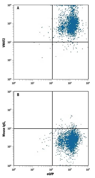
HEK293 human embryonic kidney cell line transfected with human VMAT2 and eGFP was stained with either (A) Mouse Anti-Human VMAT2 Monoclonal Antibody (Catalog # MAB8327) or (B) Mouse IgG1Isotype Control (Catalog # MAB002) followed by Allophycocyanin-conjugated Anti-Mouse IgG Secondary Antibody (Catalog # F0101B).VMAT2 in Human Brain.
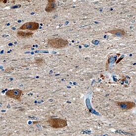
VMAT2 was detected in immersion fixed paraffin-embedded sections of human brain (substantia nigra) using Mouse Anti-Human VMAT2 Monoclonal Antibody (Catalog # MAB8327) at 15 µg/mL overnight at 4 °C. Tissue was stained using the Anti-Mouse HRP-DAB Cell & Tissue Staining Kit (brown; Catalog # CTS002) and counterstained with hematoxylin (blue). Specific staining was localized to neuronal cell bodies. View our protocol for Chromogenic IHC Staining of Paraffin-embedded Tissue Sections.Applications for Human VMAT2 Antibody
CyTOF-ready
Flow Cytometry
Sample: HEK293 human embryonic kidney cell line transfected with human VMAT2 and eGFP
Immunohistochemistry
Sample: Immersion fixed paraffin-embedded sections of human brain (substantia nigra)
Western Blot
Sample: HEK293 human embryonic kidney cell line either mock transfected or transfected with human VMAT2
Formulation, Preparation, and Storage
Purification
Reconstitution
Formulation
Shipping
Stability & Storage
- 12 months from date of receipt, -20 to -70 °C as supplied.
- 1 month, 2 to 8 °C under sterile conditions after reconstitution.
- 6 months, -20 to -70 °C under sterile conditions after reconstitution.
Background: VMAT2
The Vesicular Monoamine Transporter 2 (VMAT2), also known as VAT2 and SCL18A, is a 55-75 kDa member of the vesicular transporter family, a major facilitator superfamily. VMAT2 is a 12 transmembrane (TM) glycoprotein that is found in the membrane of brain neurosecretory vesicles. It transports monoamines (dopamine, serotonin, and particularly histamine) from the cytosol into secretion vesicles by exchanging two H+ ions for one molecule of amine. Human VMAT2 is 514 amino acids (aa) in length. It contains two cytoplasmic domains, a 20 aa and a 52 aa N- and C-terminal respectively, plus an extended 88 aa luminal loop between aa 42-129. There is one luminal, intrachain disulfide bond that contributes to amine transport (C126-C333). In addition, residues in TM domains 5-8 (aa 220-352) and 9-12 (aa 358-462) also contribute to high affinity ligand interaction. VMAT2 is constitutively phosphorylated by CKII on S511 and S513. Within the cytoplasmic C-terminus, human VMAT2 is 94% aa identical to rat VMAT2.
Long Name
Alternate Names
Gene Symbol
UniProt
Additional VMAT2 Products
Product Documents for Human VMAT2 Antibody
Product Specific Notices for Human VMAT2 Antibody
For research use only

