Human vWF-A2 Antibody
R&D Systems, part of Bio-Techne | Catalog # MAB11543

Key Product Details
Species Reactivity
Applications
Label
Antibody Source
Product Specifications
Immunogen
Asp1498-Val1665
Accession # P04275
Specificity
Clonality
Host
Isotype
Scientific Data Images for Human vWF-A2 Antibody
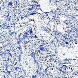
Detection of vWF-A2 in Human Placenta.
vWF-A2 was detected in immersion fixed paraffin-embedded sections of human placenta using Mouse Anti-Human vWF-A2 Monoclonal Antibody (Catalog # MAB11543) at 25 µg/ml for 1 hour at room temperature followed by incubation with the Anti-Mouse IgG VisUCyte™ HRP Polymer Antibody (Catalog # VC001) or the HRP-conjugated Anti-Mouse IgG Secondary Antibody (Catalog # HAF007). Before incubation with the primary antibody, tissue was subjected to heat-induced epitope retrieval using VisUCyte Antigen Retrieval Reagent-Basic (Catalog # VCTS021). Tissue was stained using DAB (brown) and counterstained with hematoxylin (blue). Specific staining was localized to the cytoplasm of endothelial cells. View our protocol for Chromogenic IHC Staining of Paraffin-embedded Tissue Sections.Applications for Human vWF-A2 Antibody
Immunohistochemistry
Sample: Immersion fixed paraffin-embedded sections of human placenta
Formulation, Preparation, and Storage
Purification
Reconstitution
Formulation
Shipping
Stability & Storage
- 12 months from date of receipt, -20 to -70 °C as supplied.
- 1 month, 2 to 8 °C under sterile conditions after reconstitution.
- 6 months, -20 to -70 °C under sterile conditions after reconstitution.
Background: vWF-A2
von Willebrand Factor (vWF) is a large, multimeric glycoprotein made by endothelial cells and megakaryocytes. The pre-pro-vWF protein contains 2813 amino acids (aa), which consists of 22 aa signal peptide, 741 aa propeptide, and mature vWF monomer of 2050 aa (1-4). The pro-vWF undergoes dimerization in the endoplasmic reticulum (ER) through C-terminal “cysteine-knot” (CK) domain. The pro-vWF dimmers are transported to Golgi and form multimers by forming disulfide bond in amino-terminal region of the mature form. The proteolytic processing of pro-region also occurs in Golgi. The matured vWF is stored in Weibel-Pallade bodies in endothelial cells and granules in megakaryocytes and platelets. The unusually-large vWF (ulvWF) multimers released from cells are very efficient in binding to platelets to form thrombus. The population of these highly active ulvWF multimers is controlled by a specific protease, ADAMTS13, which cleaves between residues Tyr1605 and Met1606 in the A2 domain of vWF. In the plasma, vWF appears as a series of large and intermediate multimers with molecular masses from several thousand to 500 kDa. vWF also performs hemostatic functions (3-5). In a high shear-stressed environment, vWF undergoes conformational change to expose a binding site for glycoprotein Ib alpha. As a result, vWF facilitates aggregation of platelets. In addition to platelet binding, vWF binds coagulation factor VIII to increase the lifetime of FVIII in plasma. The purified rhvWF-A2 contains the A2 domain of vWF.
References
- Sadler, J.E. (1998) Annu. Rev. Biochem. 67:395.
- Ruggeri, Z.M. (2003) Cur. Opin. Hemat. 10:142.
- Michiels, J.J. et al. (2006) Clin. Appl. Thromb. Hemost. 12:397.
- Groot, E. et al. (2007) Cur. Opin. Hemat. 14:284.
- Lenting, P.J. et al. (2007) J. Thromb. Haemos. 5:1353.
Long Name
Alternate Names
Gene Symbol
UniProt
Additional vWF-A2 Products
Product Documents for Human vWF-A2 Antibody
Product Specific Notices for Human vWF-A2 Antibody
For research use only
