Human WT1 Antibody
R&D Systems, part of Bio-Techne | Catalog # MAB57291


Conjugate
Catalog #
Key Product Details
Species Reactivity
Human
Applications
Intracellular Staining by Flow Cytometry
Label
Unconjugated
Antibody Source
Monoclonal Mouse IgG1 Clone # 960525
Product Specifications
Immunogen
E.coli-derived recombinant human WT1
Val195-Thr317
Accession # P19544
Val195-Thr317
Accession # P19544
Specificity
Detects human WT1 in direct ELISAs.
Clonality
Monoclonal
Host
Mouse
Isotype
IgG1
Scientific Data Images for Human WT1 Antibody
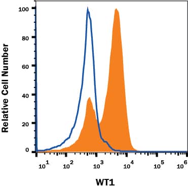
Detection of WT1 in K562 Human Cell Line by Flow Cytometry.
K562 Human chronic myelogenous leukemia cell line was stained with Mouse Anti-Human WT1 Monoclonal Antibody (Catalog # MAB57291, filled histogram) or Mouse IgG1Isotype Control Antibody (Catalog # MAB002, open histogram), followed by Allophycocyanin-conjugated Anti-Mouse IgG Secondary Antibody (Catalog # F0101B). To facilitate intracellular staining, cells were fixed and permeabilized with FlowX FoxP3 Fixation & Permeabilization Buffer Kit (Catalog # FC012). View our protocol for Staining Intracellular Molecules.Applications for Human WT1 Antibody
Application
Recommended Usage
Intracellular Staining by Flow Cytometry
0.25 µg/106 cells
Sample: K562 Human Chronic Myelogenous Leukemia Cell Line fixed and permeabilized with FlowX FoxP3 Fixation & Permeabilization Buffer Kit (Catalog # FC012)
Sample: K562 Human Chronic Myelogenous Leukemia Cell Line fixed and permeabilized with FlowX FoxP3 Fixation & Permeabilization Buffer Kit (Catalog # FC012)
Formulation, Preparation, and Storage
Purification
Protein A or G purified from hybridoma culture supernatant
Reconstitution
Reconstitute at 0.5 mg/mL in sterile PBS. For liquid material, refer to CoA for concentration.
Formulation
Lyophilized from a 0.2 μm filtered solution in PBS with Trehalose. *Small pack size (SP) is supplied either lyophilized or as a 0.2 µm filtered solution in PBS.
Shipping
Lyophilized product is shipped at ambient temperature. Liquid small pack size (-SP) is shipped with polar packs. Upon receipt, store immediately at the temperature recommended below.
Stability & Storage
Use a manual defrost freezer and avoid repeated freeze-thaw cycles.
- 12 months from date of receipt, -20 to -70 °C as supplied.
- 1 month, 2 to 8 °C under sterile conditions after reconstitution.
- 6 months, -20 to -70 °C under sterile conditions after reconstitution.
Background: WT1
Long Name
Wilms Tumor 1
Alternate Names
GUD, WAGR, WIT-2, WT33
Gene Symbol
WT1
UniProt
Additional WT1 Products
Product Documents for Human WT1 Antibody
Product Specific Notices for Human WT1 Antibody
For research use only
Loading...
Loading...
Loading...
Loading...
Loading...