Human ZAP70 Antibody
R&D Systems, part of Bio-Techne | Catalog # AF3709

Key Product Details
Species Reactivity
Validated:
Cited:
Applications
Validated:
Cited:
Label
Antibody Source
Product Specifications
Immunogen
Trp163-Cys254
Accession # P43403
Specificity
Clonality
Host
Isotype
Scientific Data Images for Human ZAP70 Antibody
Detection of Human ZAP70 by Western Blot.
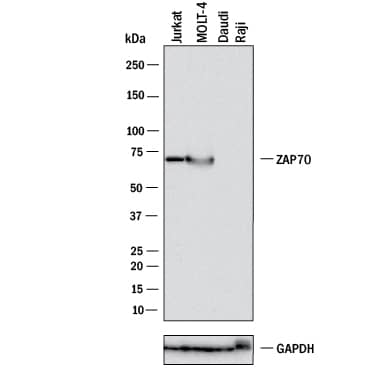
Western blot shows lysates of Jurkat human acute T cell leukemia cell line, MOLT-4 human acute lymphoblastic leukemia cell line, Daudi human Burkitt's lymphoma cell line (negative control), and Raji human Burkitt's lymphoma cell line (negative control). PVDF membrane was probed with 1 µg/mL of Goat Anti-Human ZAP70 Antigen Affinity-purified Polyclonal Antibody (Catalog # AF3709) followed by HRP-conjugated Anti-Goat IgG Secondary Antibody (HAF017). A specific band was detected for ZAP70 at approximately 70 kDa (as indicated). GAPDH is shown as a loading control. This experiment was conducted under reducing conditions and using Western Blot Buffer Group 1.ZAP70 in Human Lymphoma.
ZAP70 was detected in immersion fixed paraffin-embedded sections of human lymphoma using 10 µg/mL Human ZAP70 Antigen Affinity-purified Polyclonal Antibody (Catalog # AF3709) overnight at 4 °C. Before incubation with the primary antibody tissue was subjected to heat-induced epitope retrieval using Antigen Retrieval Reagent-Basic (Catalog # CTS013). Tissue was stained with the Anti-Goat HRP-DAB Cell & Tissue Staining Kit (brown; Catalog # CTS008) and counterstained with hematoxylin (blue). Specific labeling was localized to the cytoplasm in epithelial cells. View our protocol for Chromogenic IHC Staining of Paraffin-embedded Tissue Sections.Detection of Human ZAP70 by Simple WesternTM.
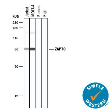
Simple Western lane view shows lysates of Jurkat human acute T cell leukemia cell line, MOLT-4 human acute lymphoblastic leukemia cell line, Ramos human Burkitt's lymphoma cell line, and Raji human Burkitt's lymphoma cell line, loaded at 0.2 mg/mL. A specific band was detected for ZAP70 at approximately 66 and 149 kDa (as indicated) using 10 µg/mL of Goat Anti-Human ZAP70 Antigen Affinity-purified Polyclonal Antibody (Catalog # AF3709) followed by 1:50 dilution of HRP-conjugated Anti-Goat IgG Secondary Antibody (Catalog # HAF109). This experiment was conducted under reducing conditions and using the 12-230 kDa separation system.Applications for Human ZAP70 Antibody
Immunohistochemistry
Sample: Immersion fixed paraffin-embedded sections of human lymphoma subjected to Antigen Retrieval Reagent-Basic (Catalog # CTS013)
Simple Western
Sample: Jurkat human acute T cell leukemia cell line and MOLT‑4 human acute lymphoblastic leukemia cell line
Western Blot
Sample: Jurkat human acute T cell leukemia cell line, and MOLT-4 human acute lymphoblastic leukemia cell line
Formulation, Preparation, and Storage
Purification
Reconstitution
Formulation
Shipping
Stability & Storage
- 12 months from date of receipt, -20 to -70 °C as supplied.
- 1 month, 2 to 8 °C under sterile conditions after reconstitution.
- 6 months, -20 to -70 °C under sterile conditions after reconstitution.
Background: ZAP70
ZAP70 (zeta-chain (TCR) associated protein kinase 70 kDa), expressed primarily in T and NK cells, is a Syk family cytosolic protein tyrosine kinase that consists of two N-terminal SH2 domains and a C-terminal tyrosine kinase domain. Upon T cell receptor activation and phosphorylation of TCR ITAMs by Src family kinases, ZAP70 is recruited to phosphorylated ITAM sequences and subsequently phosphorylated on several tyrosine residues. ZAP70 has been implicated in several immune disorders. An autosomal recessive form of SCID in humans has been attributed to a homozygous mutation in the kinase domain of ZAP70. ZAP70 expression also defines an aggressive subset of CLL.
Long Name
Alternate Names
Gene Symbol
UniProt
Additional ZAP70 Products
Product Documents for Human ZAP70 Antibody
Product Specific Notices for Human ZAP70 Antibody
For research use only


