Mouse ACE-2 Antibody
R&D Systems, part of Bio-Techne | Catalog # MAB34372


Key Product Details
Species Reactivity
Validated:
Cited:
Applications
Validated:
Cited:
Label
Antibody Source
Product Specifications
Immunogen
Gln18-Thr740
Accession # NP_081562
Specificity
Clonality
Host
Isotype
Scientific Data Images for Mouse ACE-2 Antibody
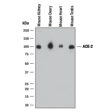
Detection of Mouse ACE-2 by Western Blot.
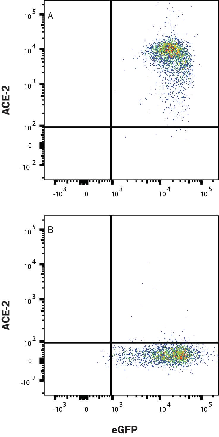
Western blot shows lysates of mouse kidney, mouse ovary, mouse heart, and mouse testis. PVDF membrane was probed with 1 µg/mL of Rabbit Anti-Mouse ACE-2 Monoclonal Antibody (Catalog # MAB34372) followed by HRP-conjugated Anti-Rabbit IgG Secondary Antibody (HAF008). A specific band was detected for ACE-2 at approximately 100 kDa (as indicated). This experiment was conducted under reducing conditions and using Western Blot Buffer Group 1.Detection of ACE-2 in HEK293 Human Cell Line Transfected with Mouse ACE-2 and eGFP by Flow Cytometry.
HEK293 human embryonic kidney cell line transfected with (A) mouse ACE-2 or (B) irrelevant protein, and eGFP was stained with Rabbit Anti-Mouse ACE-2 Monoclonal Antibody (Catalog # MAB34372) followed by Allophycocyanin-conjugated Anti-Rabbit IgG Secondary Antibody (F0111). Quadrant markers were set based on Rabbit IgG Isotype Control (MAB1050). Staining was performed using our Staining Membrane-associated Proteins protocol.ACE-2 in HEK293 Human Cell Line transfected with ACE-2.
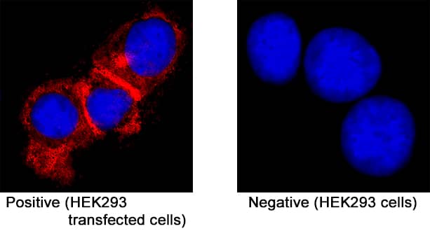
ACE-2 was detected in immersion fixed HEK293 human embryonic kidney cell line transfected with ACE-2 (positive staining) and HEK293 human embryonic kidney cell line (non-transfected, negative staining) using Rabbit Anti-Mouse ACE-2 Monoclonal Antibody (Catalog # MAB34372) at 3 µg/mL for 3 hours at room temperature. Cells were stained using the NorthernLights™ 557-conjugated Anti-Rabbit IgG Secondary Antibody (red; NL004) and counterstained with DAPI (blue). Specific staining was localized to cytoplasm. Staining was performed using our protocol for Fluorescent ICC Staining of Non-adherent Cells.Applications for Mouse ACE-2 Antibody
Flow Cytometry
Sample: HEK293 Human Cell Line Transfected with Mouse ACE-2 and eGFP
Immunocytochemistry
Sample: Immersion fixed HEK293 human embryonic kidney cell line transfected with ACE-2
Western Blot
Sample: Mouse kidney, mouse ovary, mouse heart, and mouse testis
Formulation, Preparation, and Storage
Purification
Reconstitution
Formulation
Shipping
Stability & Storage
- 12 months from date of receipt, -20 to -70 °C as supplied.
- 1 month, 2 to 8 °C under sterile conditions after reconstitution.
- 6 months, -20 to -70 °C under sterile conditions after reconstitution.
Background: ACE-2
ACE-2, also called ACEH (ACE homologue), is an integral membrane protein and a zinc metalloprotease of the ACE family that also includes somatic and germinal ACE (1). Mouse ACE-2 has about 40% amino acid identity to the N- and C-terminal domains of mouse somatic ACE. The predicted mouse ACE-2 protein sequence consists of 798 amino acids, including a N-terminal signal peptide, a single catalytic domain, a C-terminal membrane anchor, and a short cytoplasmic tail. ACE-2 cleaves angiotensins I and II as a carboxypeptidase. ACE-2 mRNA is found at high levels in testis, kidney and heart and at moderate levels in colon, small intestine and ovary. Classical ACE inhibitors such as captopril and lisinopril do not inhibit ACE-2 activity. Novel peptide inhibitors of ACE-2 do not inhibit ACE activity (2). Genetic data from Drosophila, mice and rats show that ACE-2 is an essential regulator of heart function in vivo (3). In addition, ACE-2 is a key SARS-CoV Spike protein receptor in vivo and has a critical function in acute lung injury (4, 5).
References
- Tipnis, S.R. et al. (2000) J. Biol. Chem. 275:33238.
- Crackower, M.A. et al. (2002) Nature 417:822.
- Huang, L. et al. (2003) J. Biol. Chem. 278:15532.
- Kuba, K. et al. (2005) Nature Med. 11:875.
- Ima, Y. et al. (2005) Nature 436:112.
Long Name
Alternate Names
Entrez Gene IDs
Gene Symbol
UniProt
Additional ACE-2 Products
Product Documents for Mouse ACE-2 Antibody
Product Specific Notices for Mouse ACE-2 Antibody
For research use only


