Mouse B7-H3 Antibody
R&D Systems, part of Bio-Techne | Catalog # AF1397


Key Product Details
Species Reactivity
Validated:
Cited:
Applications
Validated:
Cited:
Label
Antibody Source
Product Specifications
Immunogen
Val29-Phe244
Accession # Q8VE98
Specificity
Clonality
Host
Isotype
Endotoxin Level
Scientific Data Images for Mouse B7-H3 Antibody
Detection of Mouse B7‑H3 by Western Blot.
Western blot shows lysates of MEF mouse embryonic feeder cells, P19 mouse embryonal carcinoma cell line, NIH-3T3 mouse embryonic fibroblast cell line, C2C12 mouse myoblast cell line, and 3T3-L1 mouse embryonic fibroblast adipose-like cell line. PVDF membrane was probed with 1 µg/mL of Goat Anti-Mouse B7-H3 Antigen Affinity-purified Polyclonal Antibody (Catalog # AF1397) followed by HRP-conjugated Anti-Goat IgG Secondary Antibody (Catalog # HAF017). A specific band was detected for B7-H3 at approximately 45-55 kDa (as indicated). This experiment was conducted under reducing conditions and using Immunoblot Buffer Group 1.Proliferation Induced by B7‑H3 and Neutralization by Mouse B7‑H3 Antibody.
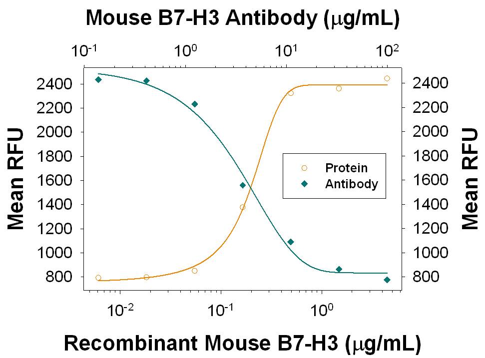
Recombinant Mouse B7-H3 enhances proliferation in mouse CD3+T cells in the presence of 100 ng/mL Hamster Anti-Mouse CD3e Monoclonal Antibody (Catalog # MAB484) in a dose-dependent manner (orange line), as measured by the Resazurin (Catalog # AR002). Proliferation elicited by Recombinant Mouse B7-H3 (2 µg/mL) is neutralized (green line) by increasing concentrations of Goat Anti-Mouse B7-H3 Antigen Affinity-purified Polyclonal Antibody (Catalog # AF1397). The ND50 is typically 1.5-7.5 µg/mL.Applications for Mouse B7-H3 Antibody
Western Blot
Sample: MEF mouse embryonic feeder cells, P19 mouse embryonal carcinoma cell line, NIH‑3T3 mouse embryonic fibroblast cell line, C2C12 mouse myoblast cell line, and 3T3‑L1 mouse embryonic fibroblast adipose-like cell line
Neutralization
Formulation, Preparation, and Storage
Purification
Reconstitution
Formulation
Shipping
Stability & Storage
- 12 months from date of receipt, -20 to -70 °C as supplied.
- 1 month, 2 to 8 °C under sterile conditions after reconstitution.
- 6 months, -20 to -70 °C under sterile conditions after reconstitution.
Background: B7-H3
T cells require a signal induced by the engagement of the T cell receptor and a “co‑stimulatory” signal(s) through distinct T cell surface molecules for optimal T cell expansion and activation. Members of the B7 superfamily of counter-receptors were identified by their ability to interact with co‑stimulatory molecules found on the surface of T cells. Members of the B7 superfamily include B7-1 (CD80), B7-2 (CD86), B7‑H1 (PD-L1), B7-H2 (B7RP-1), B7-H3, and PD-L2 (1). B7-H3 is expressed at very high levels in immature dendritic cells at moderate levels on mature dendritic cells, LPS stimulated immature dendritic cells and LPS stimulated monocytes, and at low levels on resting monocytes. B7-H3 binds to activated T cells via an as-of-yet identified receptor. B7-H3 co-stimulates proliferation of T cells and interferon-gamma (IFN-gamma) production and enhances the induction of cytotoxic T cells. B7-H3 shares 20‑27% amino acid (aa) identity with other B7 family members (2). Murine B7-H3 is a 259 aa protein containing an extracellular domain, a transmembrane domain and a cytoplasmic domain. Mouse and human B7-H3 share 87% aa identity (3).
References
- Coyle, A.J. and J.-C. Gutierrez-Ramos (2001) Nature Immunol. 2:203.
- Chapoval, A.I. et al. (2001) Nature Immunol. 2:269.
- Sun, M. et al. (2002) J. Immunol. 168:6294.
Long Name
Alternate Names
Gene Symbol
UniProt
Additional B7-H3 Products
Product Documents for Mouse B7-H3 Antibody
Product Specific Notices for Mouse B7-H3 Antibody
For research use only
