Mouse C4.4A/LYPD3 Antibody
R&D Systems, part of Bio-Techne | Catalog # AF5567

Key Product Details
Species Reactivity
Validated:
Cited:
Applications
Validated:
Cited:
Label
Antibody Source
Product Specifications
Immunogen
Leu33-His287
Accession # Q91YK8
Specificity
Clonality
Host
Isotype
Scientific Data Images for Mouse C4.4A/LYPD3 Antibody
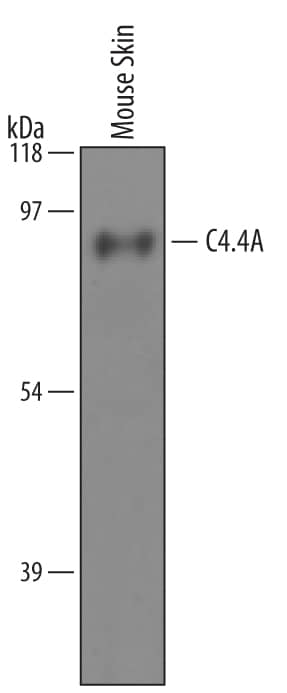
Detection of Mouse C4.4A/LYPD3 by Western Blot.
Western blot shows lysates of mouse skin tissue. PVDF membrane was probed with 1 µg/mL of Goat Anti-Mouse C4.4A/LYPD3 Antigen Affinity-purified Polyclonal Antibody (Catalog # AF5567) followed by HRP-conjugated Anti-Goat IgG Secondary Antibody (Catalog # HAF019). A specific band was detected for C4.4A/LYPD3 at approximately 95 kDa (as indicated). This experiment was conducted under reducing conditions and using Immunoblot Buffer Group 8.C4.4A/LYPD3 in Mouse Ovary.
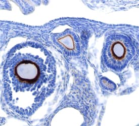
C4.4A/LYPD3 was detected in perfusion fixed frozen sections of mouse ovary using Goat Anti-Mouse C4.4A/LYPD3 Antigen Affinity-purified Polyclonal Antibody (Catalog # AF5567) at 15 µg/mL overnight at 4 °C. Tissue was stained using the Anti-Goat HRP-DAB Cell & Tissue Staining Kit (brown; Catalog # CTS008) and counterstained with hematoxylin (blue). View our protocol for Chromogenic IHC Staining of Frozen Tissue Sections.Detection of Human C4.4A/LYPD3 by Immunohistochemistry
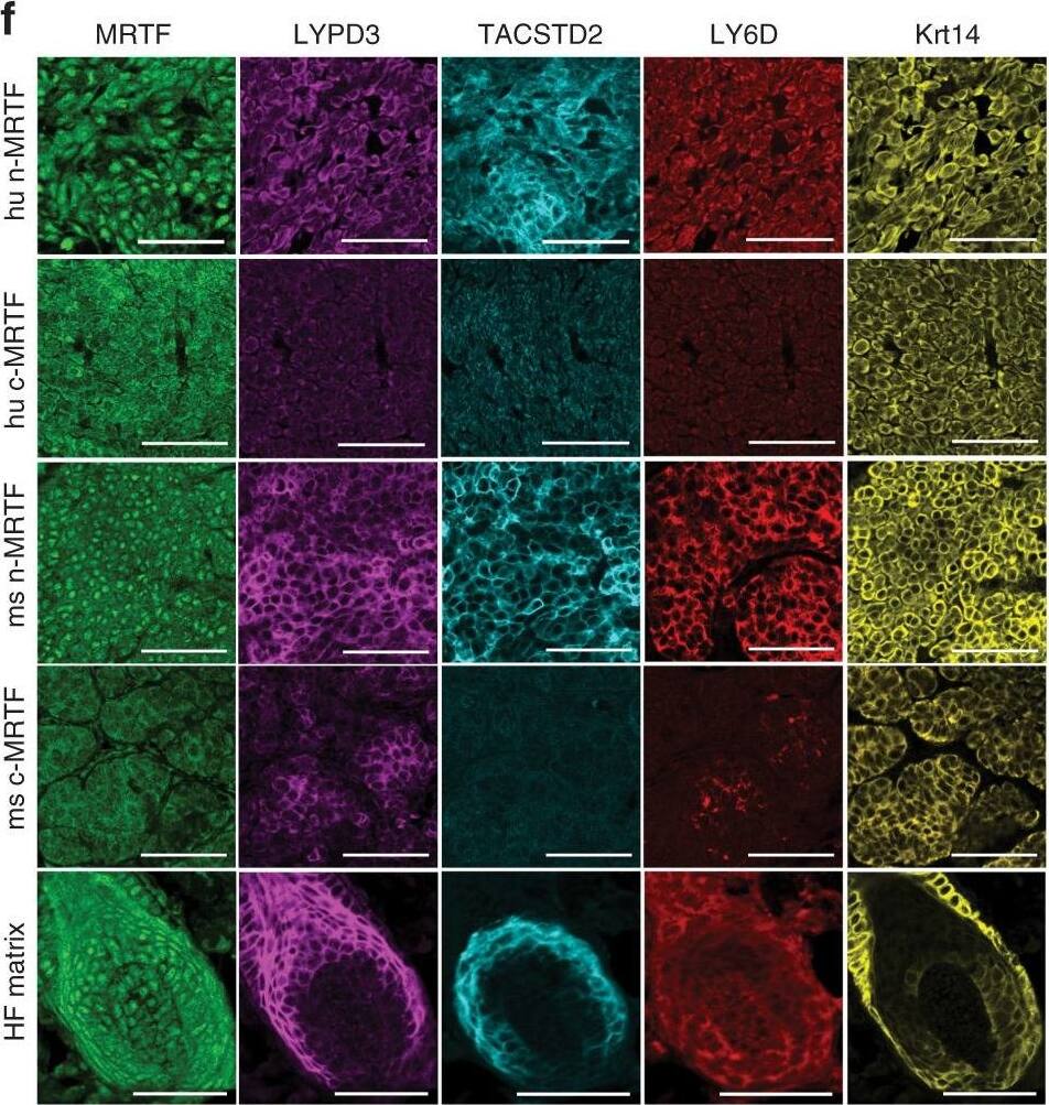
LYPD3/TACSTD2/LY6D mark the nMRTF subpopulation in patient BCCs.f Representative IF images of surface marker expression in human&mouse MRTF-nuclear vs. MRTF-cytoplasmic BCC regions or HF matrix of normal mouse skin. scale bar = 50 μm. Image collected & cropped by CiteAb from the following open publication (https://pubmed.ncbi.nlm.nih.gov/33033234), licensed under a CC-BY license. Not internally tested by R&D Systems.Applications for Mouse C4.4A/LYPD3 Antibody
Immunohistochemistry
Sample: Perfusion fixed frozen sections of mouse ovary (developing oocytes)
Western Blot
Sample: Mouse skin tissue
Formulation, Preparation, and Storage
Purification
Reconstitution
Formulation
Shipping
Stability & Storage
- 12 months from date of receipt, -20 to -70 °C as supplied.
- 1 month, 2 to 8 °C under sterile conditions after reconstitution.
- 6 months, -20 to -70 °C under sterile conditions after reconstitution.
Background: C4.4A/LYPD3
C4.4A, also known as Ly6/PLAUR domain containing 3 (LYPD-3), is a GPI-linked protein with structural similarity to the urokinase-type plasminogen activator receptor (uPAR) (1). Mature mouse C4.4A contains two uPAR/Ly6 domains and a Ser/Thr/Pro-rich (STP) region that includes a protease sensitive site (2, 3). Mouse C4.4A shares 80% and 92% amino acid sequence identity with human and rat C4.4A, respectively. It is a 65-100 kDa molecule with cell type-specific N- and O-linked glycosylation (4, 5). Proteolytic cleavage following the second uPAR/Ly6 domain generates a 35-40 kDa soluble form, while ADAM10 or ADAM17-mediated cleavage within the STP region generates a 90 kDa soluble form (6-8). Soluble C4.4A can also be shed and released in membrane vesicles (5). C4.4A is expressed in the suprabasal layers of stratified squamous epithelium and is upregulated on migrating keratinocytes during wound healing (6, 7). Its expression is downregulated during the onset of epithelial dysplasia but subsequently upregulated at the invasive front of melanomas and various carcinomas (2, 6, 5, 9). Metastases derived from these tumors also express high levels of C4.4A (2, 5, 6). The interaction of C4.4A with Laminin-1 and -5 on neighboring cells promotes the adhesion, spreading, and migration of tumor cells (4, 6, 10). C4.4A additionally interacts with Galectin-3 and the anterior gradient proteins AG-2 and AG-3 (10, 11). C4.4A over-expression in non-small cell lung cancer is predictive of increased mortality (12).
References
- Jacobsen, B. and M. Ploug (2008) Curr. Med. Chem. 15:2559.
- Smith, B.A. et al. (2001) Cancer Res. 61:1678.
- Wurfel, J. et al. (2001) Gene 262:35.
- Rosel, M. et al. (1998) Oncogene 17:1989.
- Paret, C. et al. (2007) Br. J. Cancer 97:1146.
- Hansen, L.V. et al. (2008) Int. J. Cancer 122:734.
- Hansen, L.V. et al. (2004) Biochem. J. 380:845.
- Esselens, C.W. et al. (2008) Biol. Chem. 389:1075.
- Seiter, S. et al. (2001) J. Invest. Dermatol. 116:344.
- Paret, C. et al. (2005) Int. J. Cancer 115:724.
- Fletcher, G.C. et al. (2003) Br. J. Cancer 88:579.
- Hansen, L.V. et al. (2007) Lung Cancer 58:260.
Long Name
Alternate Names
Gene Symbol
UniProt
Additional C4.4A/LYPD3 Products
Product Documents for Mouse C4.4A/LYPD3 Antibody
Product Specific Notices for Mouse C4.4A/LYPD3 Antibody
For research use only


