Mouse CCL3/MIP-1 alpha Antibody
R&D Systems, part of Bio-Techne | Catalog # AB-450-NA

Key Product Details
Validated by
Species Reactivity
Validated:
Cited:
Applications
Validated:
Cited:
Label
Antibody Source
Product Specifications
Immunogen
Ala24-Ala92
Accession # Q5QNW0
Specificity
Clonality
Host
Isotype
Endotoxin Level
Scientific Data Images for Mouse CCL3/MIP-1 alpha Antibody
Chemotaxis Induced by CCL3/MIP‑1 alpha and Neutrali-zation by Mouse CCL3/MIP‑1 alpha Antibody.
Recombinant Mouse CCL3/MIP-1a (Catalog # 450-MA) chemoattracts BaF3 mouse pro-B cell line transfected with human CCR5 in a dose-dependent manner (orange line). The amount of cells that migrated through to the lower chemotaxis chamber was measured by Resazurin (Catalog # AR002). Chemotaxis elicited by Recombinant Mouse CCL3/MIP-1a (10 ng/mL) is neutralized (green line) by increasing concentrations of Goat Anti-Mouse CCL3/MIP-1a Polyclonal Antibody (Catalog # AB-450-NA). The ND50 is typically 0.3-1 µg/mL.Detection of Mouse CCL3/MIP-1 alpha by Immunocytochemistry/ Immunofluorescence
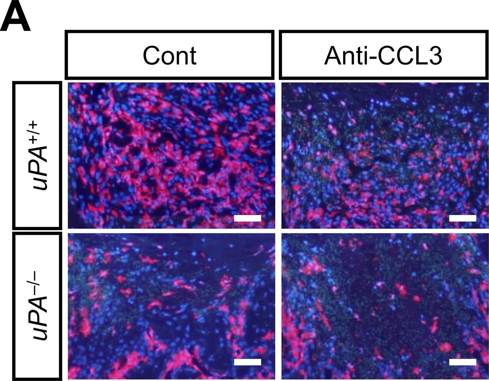
Effects of neutralizing anti-CCL3 antibody on the number of macrophages at the damaged site and the decrease in area of bone defect on day 4 in uPA+/+ and uPA-/- mice.(A) Microphotographs of immunostaining for F4/80 at the damaged site on day 4 after a femoral bone defect in uPA+/+ and uPA-/- mice treated with normal IgG (Cont) or neutralizing anti-CCL3 antibody (Anti-CCL3). The results represent experiments performed on 5 mice in each group. Scale bars indicate 50 μm. (B) Quantification of the number of F4/80-positive cells per 0.1 mm2 in the microscopic fields in the damaged site on day 4 after a femoral bone defect in uPA+/+ and uPA-/- mice treated with normal IgG or neutralizing anti-CCL3 antibody. The data represent the mean ± SEM of 5 mice. (C) Quantification of the bone defect area as assessed by qCT on days 1 and 4 in uPA+/+ and uPA-/- mice treated with normal IgG or neutralizing anti-CCL3 antibody. The data represent the mean ± SEM from 5 mice. **P < 0.01 and *P < 0.05 (Tukey’s test). Image collected and cropped by CiteAb from the following open publication (https://dx.plos.org/10.1371/journal.pone.0123982), licensed under a CC-BY license. Not internally tested by R&D Systems.Detection of Mouse CCL3/MIP-1 alpha by Immunocytochemistry/ Immunofluorescence
Effects of neutralizing anti-CCL3 antibody on the number of macrophages at the damaged site and the decrease in area of bone defect on day 4 in uPA+/+ and uPA-/- mice.(A) Microphotographs of immunostaining for F4/80 at the damaged site on day 4 after a femoral bone defect in uPA+/+ and uPA-/- mice treated with normal IgG (Cont) or neutralizing anti-CCL3 antibody (Anti-CCL3). The results represent experiments performed on 5 mice in each group. Scale bars indicate 50 μm. (B) Quantification of the number of F4/80-positive cells per 0.1 mm2 in the microscopic fields in the damaged site on day 4 after a femoral bone defect in uPA+/+ and uPA-/- mice treated with normal IgG or neutralizing anti-CCL3 antibody. The data represent the mean ± SEM of 5 mice. (C) Quantification of the bone defect area as assessed by qCT on days 1 and 4 in uPA+/+ and uPA-/- mice treated with normal IgG or neutralizing anti-CCL3 antibody. The data represent the mean ± SEM from 5 mice. **P < 0.01 and *P < 0.05 (Tukey’s test). Image collected and cropped by CiteAb from the following open publication (https://dx.plos.org/10.1371/journal.pone.0123982), licensed under a CC-BY license. Not internally tested by R&D Systems.Applications for Mouse CCL3/MIP-1 alpha Antibody
Western Blot
Sample: Recombinant Mouse CCL3/MIP-1 alpha Isoform LD78a (Catalog # 450-MA)
Neutralization
Formulation, Preparation, and Storage
Purification
Reconstitution
Formulation
Shipping
Stability & Storage
- 12 months from date of receipt, -20 to -70 °C as supplied.
- 1 month, 2 to 8 °C under sterile conditions after reconstitution.
- 6 months, -20 to -70 °C under sterile conditions after reconstitution.
Background: CCL3/MIP-1 alpha
The macrophage inflammatory proteins 1 alpha and 1 beta, two closely related but distinct proteins, were originally co-purified from medium conditioned by a LPS-stimulated murine macrophage cell line. Mature mouse MIP-1 alpha shares approximately 77% and 70% amino acid identity with human MIP-1 alpha and mouse MIP-1 beta, respectively.
MIP‑1 proteins are expressed primarily in T cells, B cells, and monocytes after antigen or mitogen stimulation. The MIP-1 proteins are members of the beta (C-C) subfamily of chemokines.
Both MIP-1 alpha and MIP-1 beta are monocyte chemoattractants in vitro. Additionally, the MIP-1 proteins have been reported to have chemoattractant and adhesive effects on lymphocytes, with MIP-1 alpha and MIP-1 beta preferentially attracting CD8+ and CD4+ T cells, respectively. MIP-1 alpha has also been shown to attract B cells as well as eosinophils. MIP-1 proteins have been reported to have multiple effects on hematopoietic precursor cells and MIP-1 alpha has been identified as a stem cell inhibitory factor that can inhibit the proliferation of hematopoietic stem cells in vitro as well as in vivo. In the same assays, MIP-1 beta was reported to be much less active. The functional receptor for MIP-1 alpha has been identified as CCR1 and CCR5.
References
- Menten, P. et al. (2002) Cytokine Growth Factor Rev. 13:455.
Alternate Names
Entrez Gene IDs
Gene Symbol
UniProt
Additional CCL3/MIP-1 alpha Products
Product Documents for Mouse CCL3/MIP-1 alpha Antibody
Product Specific Notices for Mouse CCL3/MIP-1 alpha Antibody
For research use only


