Mouse CD27/TNFRSF7 Antibody
R&D Systems, part of Bio-Techne | Catalog # MAB574

Key Product Details
Species Reactivity
Validated:
Cited:
Applications
Validated:
Cited:
Label
Antibody Source
Product Specifications
Immunogen
Thr21-Arg182
Accession # P41272
Specificity
Clonality
Host
Isotype
Scientific Data Images for Mouse CD27/TNFRSF7 Antibody
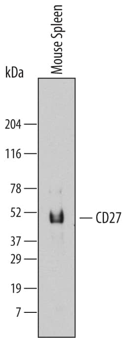
Detection of Mouse CD27/TNFRSF7 by Western Blot.
Western blot shows lysates of mouse spleen tissue. PVDF Membrane was probed with 2 µg/mL of Mouse CD27/TNFRSF7 Monoclonal Antibody (Catalog # MAB574) followed by HRP-conjugated Anti-Rat IgG Secondary Antibody (Catalog # HAF005). A specific band was detected for CD27/TNFRSF7 at approximately 45 kDa (as indicated). This experiment was conducted under reducing conditions and using Immunoblot Buffer Group 7.CD27/TNFRSF7 in Mouse Splenocytes.
CD27/TNFRSF7 was detected in immersion fixed mouse splenocytes using Rat Anti-Mouse CD27/TNFRSF7 Monoclonal Antibody (Catalog # MAB574) at 8 µg/mL for 3 hours at room temperature. Cells were stained using the NorthernLights™ 557-conjugated Anti-Rat IgG Secondary Antibody (red; Catalog # NL013) and counterstained with DAPI (blue). Specific staining was localized to plasma membrane. View our protocol for Fluorescent ICC Staining of Non-adherent Cells.Applications for Mouse CD27/TNFRSF7 Antibody
Immunocytochemistry
Sample: Immersion fixed mouse splenocytes
Western Blot
Sample: Mouse spleen tissue
Reviewed Applications
Read 1 review rated 5 using MAB574 in the following applications:
Formulation, Preparation, and Storage
Purification
Reconstitution
Formulation
Shipping
Stability & Storage
- 12 months from date of receipt, -20 to -70 °C as supplied.
- 1 month, 2 to 8 °C under sterile conditions after reconstitution.
- 6 months, -20 to -70 °C under sterile conditions after reconstitution.
Background: CD27/TNFRSF7
CD27 is a lymphocyte-specific member of the tumor necrosis factor receptor superfamily (TNFRSF) and is designated TNFRSF7 (1, 2). Mouse CD27 cDNA encodes a 250 amino acid (aa) residue type I transmembrane protein with a 20 aa putative signal peptide, a 162 aa extracellular region containing three TNFR cysteine-rich repeats, a 21 aa transmembrane domain and a 47 aa cytoplasmic region (3). Mouse and human CD27 share approximately 65% amino acid identity. CD27 exists as homodimers on the cell surface via an extracellular disulfide bond in the membrane-proximal region. A soluble form of CD27 is also produced during the immune response and is found in various body fluids (4). CD27 is expressed on subsets of T and B cells. The expression of CD27 is upregulated upon T-cell activation. Although CD27 appears to be a marker for human memory B cells, it is only expressed in a small population of mouse B cells in germinal centers and at sites of B cell stimulation, suggesting that mouse CD27 may be a marker for activated B cells (5). CD27 interacts with CD27 ligand (also named CD70 and TNFSF7), which is a member of the TNF ligand superfamily. Ligation of CD27 on T cells provides costimulatory signals that are required for T cell proliferation, clonal expansion and the promotion of effector T cell formation (1, 2). Ligation of CD27 on B cells has been shown to inhibit terminal differentiation of activated mouse B cells into plasma cells and enhances commitment to memory B cell responses (5).
References
- Croft, M. (2003) Nature Reviews Immunol. 3:609.
- Croft, M. (2003) Cytokine and Growth Factor Reviews 14:265.
- Gravestein, L.A. et al. (1993) Eur. J. Immunol. 23:943.
- Lens, S.M. et al. (1998) Semin. Immunol. 10:491.
- Raman, V.S. et al. (2003) J. Immunol. 171:5876.
Alternate Names
Gene Symbol
UniProt
Additional CD27/TNFRSF7 Products
Product Documents for Mouse CD27/TNFRSF7 Antibody
Product Specific Notices for Mouse CD27/TNFRSF7 Antibody
For research use only

