Mouse CXCR5 APC-conjugated Antibody
R&D Systems, part of Bio-Techne | Catalog # FAB6198A


Key Product Details
Species Reactivity
Validated:
Cited:
Applications
Validated:
Cited:
Label
Antibody Source
Product Specifications
Immunogen
Met1-Pro57
Accession # Q04683
Specificity
Clonality
Host
Isotype
Scientific Data Images for Mouse CXCR5 APC-conjugated Antibody
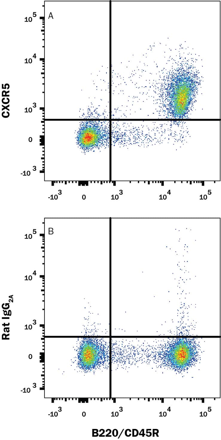
Detection of CXCR5 in Mouse Splenocytes by Flow Cytometry.
Mouse splenocytes were stained with Rat Anti-Mouse B220/CD45R PE-conjugated Monoclonal Antibody (Catalog # FAB1217P) and either (A) Rat Anti-Mouse CXCR5 APC-conjugated Monoclonal Antibody (Catalog # FAB6198A) or (B) Rat IgG2AAllophycocyanin Isotype Control (Catalog # IC006A). View our protocol for Staining Membrane-associated Proteins.Detection of CXCR5 in Mouse T Follicular Helper Cells by Flow Cytometry.
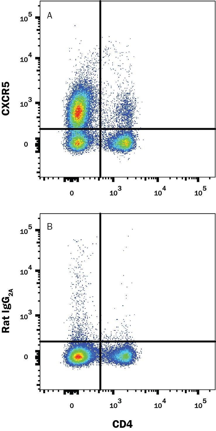
Mouse T follicular helper cells from immunized Balb/c mice were stained with Rat Anti-Mouse CD4 Alexa Fluor® 405-conjugated Monoclonal Antibody (Catalog # FAB554V) and either (A) Rat Anti-Mouse CXCR5 APC-conjugated Monoclonal Antibody (Catalog # FAB6198A) or (B) Rat IgG2AAllophycocyanin Isotype Control (Catalog # IC006A). View our protocol for Staining Membrane-associated Proteins.Detection of Human CXCR5 by Flow Cytometry
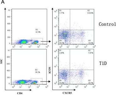
The percentages of CD4+CXCR5+ICOS5+ T cells in peripheral blood of patients with T1D.Peripheral blood mononuclear cells (PMBCs) from T1D patients (n = 54) and healthy controls (n = 31) were stained with labelled antibodies as described in Methods. A. Representative dot plots of CD4+CXCR5+ICOS5+ T cells in different groups of samples. Values in the upper right quadrant correspond to the percentages of CD4+CXCR5+ICOS5+ T cells. At least about 50,000 events were analyzed for each sample. B. CD4+CXCR5+ICOS5+ T cells were compared between T1D patients and healthy controls. Each data point represents an individual subject. The bars indicate the mean values. Student's unpaired t test was performed. ***, P<0.001. Image collected and cropped by CiteAb from the following publication (https://pubmed.ncbi.nlm.nih.gov/24278195), licensed under a CC-BY license. Not internally tested by R&D Systems.Applications for Mouse CXCR5 APC-conjugated Antibody
Flow Cytometry
Sample: Mouse splenocytes and T follicular helper cells
Formulation, Preparation, and Storage
Purification
Formulation
Shipping
Stability & Storage
- 12 months from date of receipt, 2 to 8 °C as supplied.
Background: CXCR5
CXCR5 (CXC chemokine receptor 5; also BLR-1, NLR and CD185) is a 55-60 kDa member of the G-protein coupled receptor 1 family. It is expressed on select cell types, including granule and Purkinje cell neurons, embryonic CD4+ CD3- IL-7R alpha+ precursor cells, B cells and follicular T helper cells. CXCR5 selectively binds BLC/CXCL13. This appears to both promote embryonic lymph node development and, in the adult, direct expressing cells to positions that optimize antigen presentation and antibody production. Mouse CXCR5 is a 7-transmembrane glycoprotein that is 374 amino acids (aa) in length. It contains a 57 aa N-terminal extracellular region plus a 47 aa C-terminal cytoplasmic domain. Over aa 1-57, mouse CXCR5 shares 84% and 47% aa identity with rat and human CXCR5, respectively.
Alternate Names
Gene Symbol
UniProt
Additional CXCR5 Products
Product Documents for Mouse CXCR5 APC-conjugated Antibody
Product Specific Notices for Mouse CXCR5 APC-conjugated Antibody
For research use only

