Mouse Cyr61/CCN1 Antibody
R&D Systems, part of Bio-Techne | Catalog # AF4055

Key Product Details
Species Reactivity
Validated:
Cited:
Applications
Validated:
Cited:
Label
Antibody Source
Product Specifications
Immunogen
Asp176-Gly281
Accession # NP_034646
Specificity
Clonality
Host
Isotype
Scientific Data Images for Mouse Cyr61/CCN1 Antibody
Detection of Mouse Cyr61/CCN1 by Western Blot.
Western blot shows lysates of mouse spleenocyte cell, RAW 264.7 mouse monocyte/macrophage cell line, NIH-3T3 mouse embryonic fibroblast cell line, and A20 mouse B cell lymphoma cell line. PVDF membrane was probed with 1 µg/mL of Mouse Cyr61/CCN1 Antigen Affinity-purified Polyclonal Antibody (Catalog # AF4055) followed by HRP-conjugated Anti-Sheep IgG Secondary Antibody (Catalog # HAF016). A specific band was detected for Cyr61/CCN1 at approximately 50 kDa (as indicated). This experiment was conducted under reducing conditions and using Immunoblot Buffer Group 1.Cyr61/CCN1 in Mouse Embryo.
Cyr61/CCN1 was detected in immersion fixed frozen sections of mouse embryo (13 d.p.c.) using 15 µg/mL Mouse Cyr61/CCN1 Antigen Affinity-purified Polyclonal Antibody (Catalog # AF4055) overnight at 4 °C. Tissue was stained with the Anti-Sheep HRP-DAB Cell & Tissue Staining Kit (brown; Catalog # CTS019) and counterstained with hematoxylin (blue). Specific labeling was localized to the cytoplasm of muscle cells. View our protocol for Chromogenic IHC Staining of Frozen Tissue Sections.Detection of Mouse Cyr61/CCN1 by Simple WesternTM.
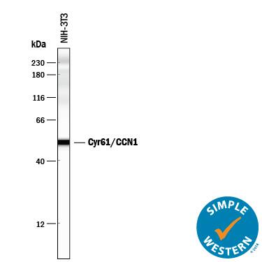
Simple Western lane view shows lysates of NIH-3T3 mouse embryonic fibroblast cell line, loaded at 0.2 mg/mL. A specific band was detected for Cyr61/CCN1 at approximately 51 kDa (as indicated) using 10 µg/mL of Sheep Anti-Mouse Cyr61/CCN1 Antigen Affinity-purified Polyclonal Antibody (Catalog # AF4055) followed by 1:50 dilution of HRP-conjugated Anti-Sheep IgG Secondary Antibody (Catalog # HAF016). This experiment was conducted under reducing conditions and using the 12-230 kDa separation system.Applications for Mouse Cyr61/CCN1 Antibody
Immunohistochemistry
Sample: Immersion fixed frozen sections of mouse embryo (13 d.p.c.)
Simple Western
Sample: NIH‑3T3 mouse embryonic fibroblast cell line
Western Blot
Sample: Mouse spleenocyte cell, RAW 264.7 mouse monocyte/macrophage cell line, NIH-3T3 mouse embryonic fibroblast cell line, and A20 mouse B cell lymphoma cell line
Reviewed Applications
Read 2 reviews rated 5 using AF4055 in the following applications:
Formulation, Preparation, and Storage
Purification
Reconstitution
Formulation
Shipping
Stability & Storage
- 12 months from date of receipt, -20 to -70 °C as supplied.
- 1 month, 2 to 8 °C under sterile conditions after reconstitution.
- 6 months, -20 to -70 °C under sterile conditions after reconstitution.
Background: Cyr61/CCN1
Cyr61, also known as IGFBP-10 and CCN1, is a 50 kDa secreted matrix- and cell-associated protein that regulates the growth and adhesion of vascular endothelial cells, fibroblasts, and monocytes. Cyr61 interacts with cells that express integrins alphaV beta3, alphaV beta5, alphaM beta2, and alpha6 beta1. Cyr61 is cleaved by plasmin within its VWF domain which generates an N-terminal fragment that is not associated with the matrix but retains the ability to induce endothelial cell migration. Cyr61 induces VEGF upregulation, angiogenesis, and tumorigenesis. Between amino acids 176‑281, mouse Cyr61 shares 87% and 97% amino acid sequence identity with human and rat Cyr61, respectively.
Long Name
Alternate Names
Gene Symbol
UniProt
Additional Cyr61/CCN1 Products
Product Documents for Mouse Cyr61/CCN1 Antibody
Product Specific Notices for Mouse Cyr61/CCN1 Antibody
For research use only


