Mouse Endomucin Antibody
R&D Systems, part of Bio-Techne | Catalog # AF4666

Key Product Details
Validated by
Species Reactivity
Validated:
Cited:
Applications
Validated:
Cited:
Label
Antibody Source
Product Specifications
Immunogen
Glu21-Glu90
Accession # Q9R0H2
Specificity
Clonality
Host
Isotype
Scientific Data Images for Mouse Endomucin Antibody
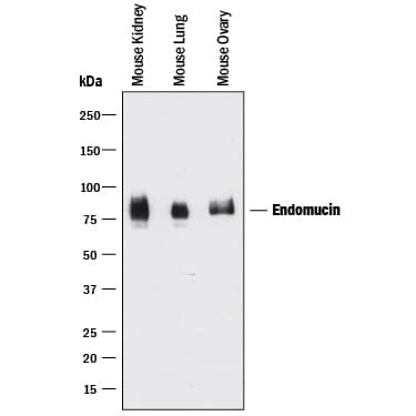
Detection of Mouse Endomucin by Western Blot.
Western blot shows lysates of mouse kidney tissue, mouse lung tissue, and mouse ovary tissue. PVDF membrane was probed with 1 µg/mL of Goat Anti-Mouse Endomucin Antigen Affinity-purified Polyclonal Antibody (Catalog # AF4666) followed by HRP-conjugated Anti-Goat IgG Secondary Antibody (Catalog # HAF017). A specific band was detected for Endomucin at approximately 75-85 kDa (as indicated). This experiment was conducted under reducing conditions and using Immunoblot Buffer Group 1.Endomucin in bEnd.3 Mouse Cell Line.
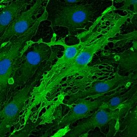
Endomucin was detected in immersion fixed bEnd.3 mouse endothelioma cell line using Goat Anti-Mouse Endomucin Antigen Affinity-purified Polyclonal Antibody (Catalog # AF4666) at 10 µg/mL for 3 hours at room temperature. Cells were stained using the NorthernLights™ 493-conjugated Anti-Goat IgG Secondary Antibody (green; Catalog # NL003) and counterstained with DAPI (blue). Specific staining was localized to cytoplasm. View our protocol for Fluorescent ICC Staining of Cells on Coverslips.Detection of Endomucin in bEnd.3 Mouse Cell Line by Flow Cytometry.
bEnd.3 mouse endothelioma cell line was stained with Goat Anti-Mouse Endomucin Antigen Affinity-purified Polyclonal Antibody (Catalog # AF4666, filled histogram) or isotype control antibody (Catalog # AB-108-C, open histogram), followed by Allophycocyanin-conjugated Anti-Goat IgG Secondary Antibody (Catalog # F0108).Applications for Mouse Endomucin Antibody
CyTOF-ready
Flow Cytometry
Sample: bEnd.3 mouse endothelioma cell line
Immunocytochemistry
Sample: Immersion fixed bEnd.3 mouse endothelioma cell line
Western Blot
Sample: Mouse kidney tissue, mouse lung tissue, and mouse ovary tissue
Reviewed Applications
Read 1 review rated 4 using AF4666 in the following applications:
Formulation, Preparation, and Storage
Purification
Reconstitution
Formulation
Shipping
Stability & Storage
- 12 months from date of receipt, -20 to -70 °C as supplied.
- 1 month, 2 to 8 °C under sterile conditions after reconstitution.
- 6 months, -20 to -70 °C under sterile conditions after reconstitution.
Background: Endomucin
Endomucin (endothelial sialomucin; also Endomucin-1/2 and Mucin-14) is an 80‑120 kDa glycoprotein member of the Endomucin family of proteins. It is expressed on endothelial cells and depending upon its glycosylation pattern, can serve as either a pro- or anti-adhesive molecule. Mouse Endomucin precursor is 261 amino acids in length. It is type I transmembrane protein that contains a 170 aa extracellular domain (ECD) (aa 21‑190) and a 50 aa cytoplasmic region. Three splice variants exist in the ECD. One shows a deletion of aa 91‑141, a second shows a one aa substitution for aa 91‑129, and a third shows a one aa substitution for aa 129‑142. Over
aa 21‑90, mouse Endomucin shares 60% and 30% aa identity with rat and human Endomucin, respectively.
Alternate Names
Gene Symbol
UniProt
Additional Endomucin Products
Product Documents for Mouse Endomucin Antibody
Product Specific Notices for Mouse Endomucin Antibody
For research use only
