Mouse GRIN1/NMDAR1 Antibody
R&D Systems, part of Bio-Techne | Catalog # MAB10655


Conjugate
Catalog #
Key Product Details
Species Reactivity
Mouse
Applications
Immunocytochemistry, Intracellular Staining by Flow Cytometry
Label
Unconjugated
Antibody Source
Monoclonal Rat IgG2A Clone # 1031915
Product Specifications
Immunogen
Mouse myeloma cell line NS0-derived recombinant mouse GRIN1/NMDAR1
Met1-Gln559
Met1-Gln559
Specificity
Detects mouse GRIN1/NMDAR1 in direct ELISAs.
Clonality
Monoclonal
Host
Rat
Isotype
IgG2A
Scientific Data Images for Mouse GRIN1/NMDAR1 Antibody
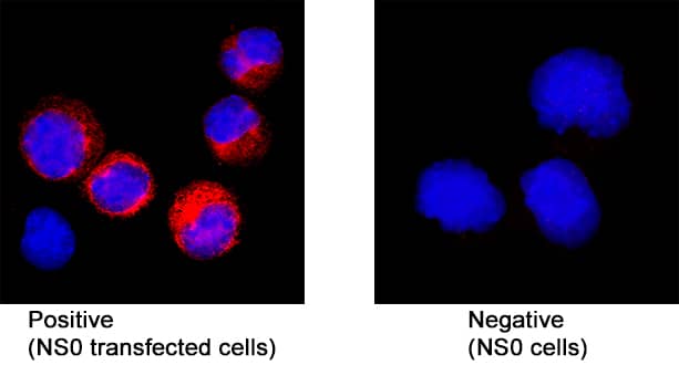
GRIN1/NMDAR1 in NS0 Mouse Cell Line Transfected with Mouse GRIN1/NMDAR1.
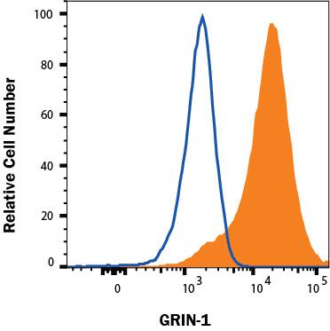
GRIN1/NMDAR1 was detected in immersion fixed NS0 mouse myeloma cell line transfected with mouse GRIN1/NMDAR1 (positive staining) and NS0 mouse myeloma cell line (wild type, negative control) using Rat Anti-Mouse GRIN1/NMDAR1 Monoclonal Antibody (Catalog # MAB10655) at 8 µg/mL for 3 hours at room temperature. Cells were stained using the NorthernLights™ 557-conjugated Anti-Rat IgG Secondary Antibody (red; NL013) and counterstained with DAPI (blue). Specific staining was localized to cytoplasm. Staining was performed using our protocol for Fluorescent ICC Staining of Non-adherent Cells.Detection of Mouse GRIN1/NMDAR1 in NS0 cells transfected with Mouse GRIN1/NMDAR1 by Flow Cytometry.
NS0 cells transfected with mouse GRIN1/NMDAR1 (filled histogram) or irrelevant protein (open histogram) were stained with Rat Anti-Mouse GRIN1/NMDAR1 Monoclonal Antibody (Catalog # MAB10655) or Rat IgG2A Isotype Control Antibody (MAB006, data not shown) followed by Allophycocyanin-conjugated Anti-Rat IgG Secondary Antibody (F0113). To facilitate intracellular staining, cells were fixed and permeabilized with FlowX FoxP3 Fixation & Permeabilization Buffer Kit (FC012).Applications for Mouse GRIN1/NMDAR1 Antibody
Application
Recommended Usage
Immunocytochemistry
8-25 µg/mL
Sample: Immersion fixed NS0 mouse myeloma cell line transfected with mouse GRIN1/NMDAR1
Sample: Immersion fixed NS0 mouse myeloma cell line transfected with mouse GRIN1/NMDAR1
Intracellular Staining by Flow Cytometry
0.25 µg/106 cells
Sample: NS0 cells transfected with Mouse GRIN1/NMDAR1
Sample: NS0 cells transfected with Mouse GRIN1/NMDAR1
Formulation, Preparation, and Storage
Purification
Protein A or G purified from cell culture supernatant
Reconstitution
Reconstitute at 0.5 mg/mL in sterile PBS. For liquid material, refer to CoA for concentration.
Formulation
Lyophilized from a 0.2 μm filtered solution in PBS with Trehalose. *Small pack size (SP) is supplied either lyophilized or as a 0.2 µm filtered solution in PBS.
Shipping
Lyophilized product is shipped at ambient temperature. Liquid small pack size (-SP) is shipped with polar packs. Upon receipt, store immediately at the temperature recommended below.
Stability & Storage
Use a manual defrost freezer and avoid repeated freeze-thaw cycles.
- 12 months from date of receipt, -20 to -70 °C as supplied.
- 1 month, 2 to 8 °C under sterile conditions after reconstitution.
- 6 months, -20 to -70 °C under sterile conditions after reconstitution.
Background: GRIN1/NMDAR1
Long Name
Glutamate Receptor, Ionotropic, N-Methyl-D-Aspartate, Subunit 1
Alternate Names
GluN1, MRD8, NMD-R1, NMDA R, NR1 Subunit, NMDA1, NMDAR1
Gene Symbol
GRIN1
Additional GRIN1/NMDAR1 Products
Product Documents for Mouse GRIN1/NMDAR1 Antibody
Product Specific Notices for Mouse GRIN1/NMDAR1 Antibody
For research use only
Loading...
Loading...
Loading...
Loading...
Loading...
