Mouse IL-10 Antibody
R&D Systems, part of Bio-Techne | Catalog # MAB417

Key Product Details
Validated by
Biological Validation
Species Reactivity
Validated:
Mouse
Cited:
Human, Mouse, Rat
Applications
Validated:
ELISA Capture (Matched Antibody Pair), Neutralization, Western Blot
Cited:
Bioassay, ELISA Capture, ELISA Development, ELISA Development (Capture), ELISpot Development, Flow Cytometry, Functional Assay, Immunohistochemistry, Immunohistochemistry-Paraffin, Luminex Development, Neutralization, Tissue Culture, Western Blot
Label
Unconjugated
Antibody Source
Monoclonal Rat IgG1 Clone # JES052A5
Product Specifications
Immunogen
E. coli-derived recombinant mouse IL-10
Accession # P18893
Accession # P18893
Specificity
Detects mouse IL-10 in sandwich ELISAs and Western blots. In sandwich ELISAs, less than 8% cross-reactivity with recombinant rat IL-10 and 1% cross-reactivity with recombinant cotton rat IL‑10 is observed. In sandwich ELISAs, no cross-reactivity with recombinant human (rh) IL-10, recombinant mouse IL-10 R, rhIL‑10 R, recombinant canine IL-10, recombinant feline IL-10, or recombinant porcine IL-10 is observed.
Clonality
Monoclonal
Host
Rat
Isotype
IgG1
Endotoxin Level
<0.10 EU per 1 μg of the antibody by the LAL method.
Scientific Data Images for Mouse IL-10 Antibody
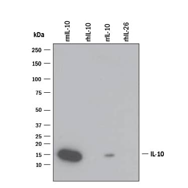
Detection of Recombinant Mouse IL‑10 by Western Blot.
Western blot shows 25 ng of Recombinant Mouse IL-10 (Catalog # 417-ML), Recombinant Human IL-10 (Catalog # 217-IL), Recombinant Rat IL-10 (Catalog # 522-RLB), and Recombinant Human IL-26/AK155 Monomer (Catalog # 1375-IL). PVDF Membrane was probed with 1 µg/mL of Rat Anti-Mouse IL-10 Monoclonal Antibody (Catalog # MAB417) followed by HRP-conjugated Anti-Rat IgG Secondary Antibody (Catalog # HAF005). A specific band was detected for IL-10 at approximately 15 kDa (as indicated). This experiment was conducted under reducing conditions and using Immunoblot Buffer Group 3.Cell Proliferation Induced by IL‑10 and Neutralization by Mouse IL‑10 Antibody.
Recombinant Mouse IL-10 (Catalog # 417-ML) stimulates proliferation in the MC/9-2 mouse mast cell line in a dose-dependent manner (orange line). Proliferation elicited by Recombinant Mouse IL-10 (2.5 ng/mL) is neutralized (green line) by increasing concen-trations of Rat Anti-Mouse IL-10 Monoclonal Antibody (Catalog # MAB417). The ND50 is typically 0.005-0.015 µg/mL.Detection of Mouse IL-10 by Western Blot
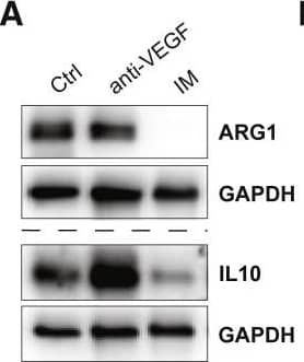
Imipramine downregulates an M2-like program in TAMs(A) Western blot analysis of ARG1 and IL-10 in single tumors treated or not with IM or anti-VEGF.(B) Flow cytometry analysis of ARG1 and IL-10 expression in GBM tumors treated for 1 week. Ctrl (n = 9 tumors), IM (n = 6), anti-VEGF (n = 6), anti-VEGFR2 (n = 6), Axitinib (n = 6). Macrophages were gated as CD45+CD11b+Ly6C-Ly6G−.(C) Expression of ARG1 and IL-10 in microglia (CD49d−) and MDMs (CD49d+) assessed by FACS in untreated tumors (n = 5).(D) Expression of MHC-II within microglia as assessed by flow cytometry. Ctrl (n = 4 tumors) and IM + anti-VEGF (n = 4).(E) Ex vivo co-cultures of tumoral CD11b cells and activated splenic CFSE-labeled CD8 or CD4 T cells. Each dot represents the average of two or three technical replicates. T cells alone (n = 4), Ctrl co-culture (n = 5), anti-VEGF (n = 3), IM (n = 4), IM + anti-VEGF (n = 4).(F) Analysis of the M2-like program in cytokine-polarized macrophages as assessed by qRT-PCR analysis of Ctrl and IM-treated M2-like BMDMs. Expression is normalized to 18S statistics by Welch’s t test. Each dot represents an individual sample. Data are representative of three independent experiments.(G) Analysis of the M1-like program in BMDMs assessed by FACS. Each dot represents an individual replicate.(H) Expression of Hrh1 mRNA normalized to 18S in ex vivo M1-and M2-polarized BMDMs, either untreated or IM treated for 24 h. Each dot represents an individual sample. Data are representative of three independent experiments.(I) mRNA expression of Hrh1 in FACS-sorted microglia or MDMs from Ctrl and IM-treated tumors.(J) mRNA expression of Arg1, Chil3, and Il10 in M2-polarized macrophages that were transfected with siCtrl or two different siHrh1 constructs. Cells were treated with 40 μm IM for 24 h. Data are representative of two independent experiments.(K) Western blot analysis of MRC1 and ARG1 expression of siRNA-transfected M2 BMDMs. Data are representative of two independent experiments.(L) CD8 and CD4 T cell proliferation during co-culture with tumoral CD11b cells isolated from untreated (n = 2) or TFP-treated tumors (n = 4).(M) mRNA expression of Hrh1, Arg1, and MMP2 in CD11b cells isolated from tumors treated with IM (n = 4), TFP (n = 4), or untreated Ctrl (n = 5).(N) Phagocytosis assay involving sorted microglia and MDMs from untreated or IM-treated tumors assayed with green pHrodo S. aureus bioparticles. Data presented as mean fluorescence intensity (MFI) of pHrodo/live cells. (Para break) Data in all quantitative panels are presented as mean ± SEM ∗p < 0.05; ∗∗p < 0.01; ∗∗∗p < 0.001; ∗∗∗∗p < 0.0001; ns, no statistical significance. Statistical analysis by Mann-Whitney test or one-way ANOVA, unless otherwise stated. Image collected and cropped by CiteAb from the following publication (https://pubmed.ncbi.nlm.nih.gov/36113478), licensed under a CC-BY license. Not internally tested by R&D Systems.Applications for Mouse IL-10 Antibody
Application
Recommended Usage
Western Blot
1 µg/mL
Sample: Recombinant Mouse IL-10 (Catalog # 417-ML)
Sample: Recombinant Mouse IL-10 (Catalog # 417-ML)
Neutralization
Measured by its ability to neutralize IL‑10-induced proliferation in the MC/9‑2 mouse mast cell line. Thompson-Snipes, L. et al. (1991) J. Exp. Med. 173:507. The Neutralization Dose (ND50) is typically 0.005-0.015 µg/mL in the presence of 2.5 ng/mL Recombinant Mouse IL‑10.
Mouse IL-10 Sandwich Immunoassay
Please Note: Optimal dilutions of this antibody should be experimentally determined.
Reviewed Applications
Read 2 reviews rated 5 using MAB417 in the following applications:
Formulation, Preparation, and Storage
Purification
Protein A or G purified from hybridoma culture supernatant
Reconstitution
Reconstitute at 0.5 mg/mL in sterile PBS. For liquid material, refer to CoA for concentration.
Formulation
Lyophilized from a 0.2 μm filtered solution in PBS with Trehalose. *Small pack size (SP) is supplied either lyophilized or as a 0.2 µm filtered solution in PBS.
Shipping
Lyophilized product is shipped at ambient temperature. Liquid small pack size (-SP) is shipped with polar packs. Upon receipt, store immediately at the temperature recommended below.
Stability & Storage
Use a manual defrost freezer and avoid repeated freeze-thaw cycles.
- 12 months from date of receipt, -20 to -70 °C as supplied.
- 1 month, 2 to 8 °C under sterile conditions after reconstitution.
- 6 months, -20 to -70 °C under sterile conditions after reconstitution.
Background: IL-10
Long Name
Interleukin 10
Alternate Names
CSIF, GVHDS, IL10, IL10A, TGIF
Entrez Gene IDs
Gene Symbol
IL10
UniProt
Additional IL-10 Products
Product Documents for Mouse IL-10 Antibody
Product Specific Notices for Mouse IL-10 Antibody
For research use only
Loading...
Loading...
Loading...
Loading...


