Mouse Notch-3 Antibody
R&D Systems, part of Bio-Techne | Catalog # AF1308


Key Product Details
Species Reactivity
Validated:
Cited:
Applications
Validated:
Cited:
Label
Antibody Source
Product Specifications
Immunogen
Ala40-Glu468
Accession # Q61982
Specificity
Clonality
Host
Isotype
Endotoxin Level
Scientific Data Images for Mouse Notch-3 Antibody
Detection of Notch-3 in CD4- CD8- mouse thymocytes by Flow Cytometry
CD4- CD8- mouse thymocytes were stained with Rat Anti-Mouse CD25/IL-2R alpha PE-conjugated Monoclonal Antibody (Catalog # FAB2438P) and either (A) Goat Anti-Mouse Notch-3 Antigen Affinity-purified Polyclonal Antibody (Catalog # AF1308) or (B) isotype control antibody (Catalog # AB-108-C) followed by Allophycocyanin-conjugated Anti-Goat IgG Secondary Antibody (Catalog # F0108). View our protocol for Staining Membrane-associated Proteins.Detection of Notch-3 in Mouse Embryo (E13).
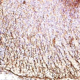
Notch-3 was detected in immersion fixed frozen sections of mouse embryo (E13) using Goat Anti-Mouse Notch-3 Antigen Affinity-purified Polyclonal Antibody (Catalog # AF1308) at 1 µg/ml for 1 hour at room temperature followed by incubation with the Anti-Goat IgG VisUCyte™ HRP Polymer Antibody (Catalog # VC004). Tissue was stained using DAB (brown) and counterstained with hematoxylin (blue). Specific staining was localized to the cell surface. View our protocol for Chromogenic IHC Staining of Frozen Tissue Sections.Detection of Notch-3 in Mouse Embryo (E13).
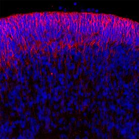
Notch-3 was detected in immersion fixed frozen sections of mouse embryo (E13) using Goat Anti-Mouse Notch-3 Antigen Affinity-purified Polyclonal Antibody (Catalog # AF1308) at 3 µg/ml overnight at 4 °C. Tissue was stained using the NorthernLights™ 557-conjugated Anti-Goat IgG Secondary Antibody (red; Catalog # NL001) and counterstained with DAPI (blue). Specific staining was localized to the cell surface. View our protocol for Fluorescent IHC Staining of Frozen Tissue Sections.Applications for Mouse Notch-3 Antibody
Blockade of Receptor-ligand Interaction
CyTOF-ready
Flow Cytometry
Sample: - D3 mouse embryonic stem cell line- CD4- CD8- mouse thymocytes
Immunofluorescence
Sample: Immersion fixed frozen sections of mouse embryo (E13)
Immunohistochemistry
Sample: Immersion fixed frozen sections of mouse embryo (E13)
Western Blot
Sample: Recombinant Mouse Notch-3 Fc Chimera, aa 40-468 (Catalog # 1308-NT)
Formulation, Preparation, and Storage
Purification
Reconstitution
Formulation
*Small pack size (-SP) is supplied either lyophilized or as a 0.2 µm filtered solution in PBS.
Shipping
Stability & Storage
- 12 months from date of receipt, -20 to -70 °C as supplied.
- 1 month, 2 to 8 °C under sterile conditions after reconstitution.
- 6 months, -20 to -70 °C under sterile conditions after reconstitution.
Background: Notch-3
Mouse Notch-3 is part of the Notch family of type I transmembrane glycoproteins involved in a number of early-event developmental processes (1). The extracellular domain of Notch receptors interact with the extracellular domain of transmembrane ligands Jagged, Delta, and Serrate expressed on the surface of a neighboring cell. In both vertebrates and invertebrates, Notch signaling is important for specifying cell fates and for defining boundaries between different cell types. The Notch molecule is synthesized as a 2318 amino acid (aa) precursor that contains an 39 aa signal sequence, a 1603 aa extracellular region, a 20 aa transmembrane (TM) segment and a 655 aa cytoplasmic domain. The large Notch extracellular domain has 34 EGF-like repeats followed by three notch/Lin-12 repeats (LNR) (2). The 11th and 12th EGF-like repeats of Notch have been shown to be both necessary and sufficient for binding the ligands Serrate and Delta, in Drosophila (3). Notch-3 has the same biochemical mechanism of signal tranduction as Notch-1, where a series of cleavage events result in the release of the Notch intracellular domain (NICD). NICD translocates into the nucleus and initiates transcription of Notch-responsive genes (4). Thus Notch acts as both a ligand-binding receptor and a nuclear factor that regulates transcription.
Notch-3 is predominantly expressed in the developing central nervous system of mice (2). Mutations in Notch-3 in humans cause an autosomal dominant condition called CADASIL (cerebral autosomal dominant arteriopathy with subcortical infarcts and leukoencephalopathy). This disorder is characterized by recurrent ischemic strokes at an early age without any underlying vascular risk and progressive dementia. Nearly all mutations leading to this disorder are clustered in the first 5 EGF repeats of the Notch-3 gene (5). Mouse Notch-3 shows 90% aa identity to human Notch-3 and 96% to rat Notch-3 over the entire protein.
References
- Weinmaster, G. (2000) Curr. Opin. Genet. Dev. 10:363.
- Lardelli, M. et al. (1994) Mech Dev. 46:123.
- Rebay, I. et al. (1991) Cell 67:687.
- Mizutani, T. et al. (2001) Proc. Natl. Acad. Sci. USA 98:9026.
- Joutel, A. and E. Tounier-Lasserve (1998) Stem Cell & Dev. Biol. 9:619.
Alternate Names
Gene Symbol
UniProt
Additional Notch-3 Products
Product Documents for Mouse Notch-3 Antibody
Product Specific Notices for Mouse Notch-3 Antibody
For research use only

