Mouse PD-1 Antibody
R&D Systems, part of Bio-Techne | Catalog # AF1021


Key Product Details
Species Reactivity
Validated:
Mouse
Cited:
Human, Mouse, Xenograft
Applications
Validated:
CyTOF-ready, Flow Cytometry, Immunohistochemistry, Western Blot
Cited:
ELISA Capture, Immunocytochemistry, Immunohistochemistry, Immunohistochemistry-Frozen, Immunohistochemistry-Paraffin, Western Blot
Label
Unconjugated
Antibody Source
Polyclonal Goat IgG
Product Specifications
Immunogen
S. frugiperda insect ovarian cell line Sf 21-derived recombinant mouse PD-1
Leu25-Gln167
Accession # Q02242
Leu25-Gln167
Accession # Q02242
Specificity
Detects mouse PD-1 in direct ELISAs and Western blots. In direct ELISAs, approximately 20% cross-reactivity with recombinant human PD‑1 is observed.
Clonality
Polyclonal
Host
Goat
Isotype
IgG
Scientific Data Images for Mouse PD-1 Antibody
Detection of Mouse PD‑1 by Western Blot.
Western blot shows lysates of EL-4 mouse lymphoblast cell line and CTLL-2 mouse cytotoxic T cell line (negative control). PVDF membrane was probed with 0.2 µg/mL of Goat Anti-Mouse PD-1 Antigen Affinity-purified Polyclonal Antibody (Catalog # AF1021) followed by HRP-conjugated Anti-Goat IgG Secondary Antibody (Catalog # HAF017). A specific band was detected for PD-1 at approximately 55 kDa (as indicated). This experiment was conducted under reducing conditions and using Immunoblot Buffer Group 1.Detection of Mouse PD‑1 by Western Blot.
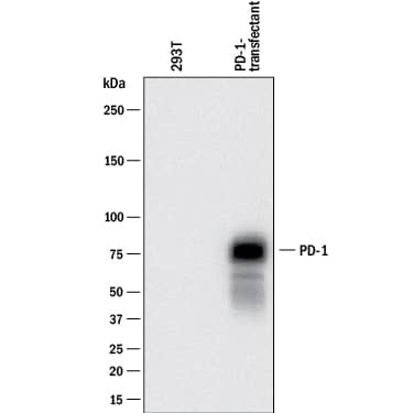
Western blot shows lysates of 293T human embryonic kidney cell line mock transfected or transfected with mouse PD-1. PVDF membrane was probed with 0.2 µg/mL of Goat Anti-Mouse PD-1 Antigen Affinity-purified Polyclonal Antibody (Catalog # AF1021) followed by HRP-conjugated Anti-Goat IgG Secondary Antibody (Catalog # HAF017). A specific band was detected for PD-1 at approximately 75 kDa (as indicated). This experiment was conducted under reducing conditions and using Immunoblot Buffer Group 1.PD-1 in Mouse Thymus.
PD-1 was detected in perfusion fixed frozen sections of mouse thymus using 15 µg/mL Mouse PD-1 Antigen Affinity-purified Polyclonal Antibody (Catalog # AF1021) overnight at 4 °C. Tissue was stained (red) and counterstained (green). View our protocol for Fluorescent IHC Staining of Frozen Tissue Sections.Applications for Mouse PD-1 Antibody
Application
Recommended Usage
CyTOF-ready
Ready to be labeled using established conjugation methods. No BSA or other carrier proteins that could interfere with conjugation.
Flow Cytometry
0.25 µg/106 cells
Sample: Mouse splenocytes treated with PHA
Sample: Mouse splenocytes treated with PHA
Immunohistochemistry
5-15 µg/mL
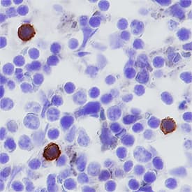
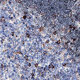
Sample: Perfusion fixed frozen sections of mouse thymus and mouse spleen, and perfusion fixed paraffin-embedded sections of mouse thymus
Sample: Perfusion fixed frozen sections of mouse thymus and mouse spleen, and perfusion fixed paraffin-embedded sections of mouse thymus
Western Blot
0.2 µg/mL
Sample: EL‑4 mouse lymphoblast cell line and 293T human embryonic kidney cell line transfected with mouse PD-1
Sample: EL‑4 mouse lymphoblast cell line and 293T human embryonic kidney cell line transfected with mouse PD-1
Reviewed Applications
Read 5 reviews rated 4.6 using AF1021 in the following applications:
Formulation, Preparation, and Storage
Purification
Antigen Affinity-purified
Reconstitution
Reconstitute at 0.2 mg/mL in sterile PBS. For liquid material, refer to CoA for concentration.
Formulation
Lyophilized from a 0.2 μm filtered solution in PBS with Trehalose. *Small pack size (SP) is supplied either lyophilized or as a 0.2 µm filtered solution in PBS.
Shipping
Lyophilized product is shipped at ambient temperature. Liquid small pack size (-SP) is shipped with polar packs. Upon receipt, store immediately at the temperature recommended below.
Stability & Storage
Use a manual defrost freezer and avoid repeated freeze-thaw cycles.
- 12 months from date of receipt, -20 to -70 °C as supplied.
- 1 month, 2 to 8 °C under sterile conditions after reconstitution.
- 6 months, -20 to -70 °C under sterile conditions after reconstitution.
Background: PD-1
References
- Ishida, Y. et al. (1992) EMBO J. 11:3887.
- Sharpe, A.H. and G.J. Freeman (2002) Nat. Rev. Immunol. 2:116.
- Coyle, A. and J. Gutierrez-Ramos (2001) Nat. Immunol. 2:203.
- Nishimura, H. and T. Honjo (2001) Trends in Immunol. 22:265.
- Latchman Y. et al. (2001) Nature Immun. 2:261.
- Tamura, H. et al. (2001) Blood 97:1809.
Long Name
Programmed Death-1
Alternate Names
CD279, PD1, PDCD1, SLEB2
Entrez Gene IDs
Gene Symbol
PDCD1
UniProt
Additional PD-1 Products
Product Documents for Mouse PD-1 Antibody
Product Specific Notices for Mouse PD-1 Antibody
For research use only
Loading...
Loading...
Loading...
Loading...
Loading...


