Mouse PU.1/Spi-1 Antibody
R&D Systems, part of Bio-Techne | Catalog # MAB7124

Key Product Details
Species Reactivity
Applications
Label
Antibody Source
Product Specifications
Immunogen
Met1-Lys169
Accession # P17433
Specificity
Clonality
Host
Isotype
Scientific Data Images for Mouse PU.1/Spi-1 Antibody
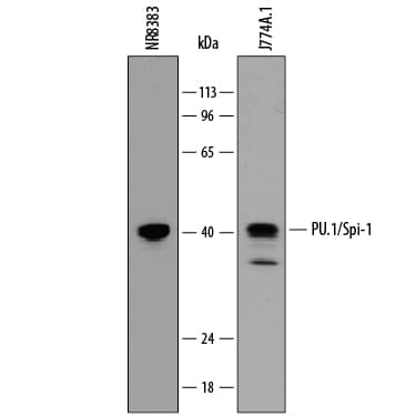
Detection of Mouse and Rat PU.1/Spi‑1 by Western Blot.
Western blot shows lysates of NR8383 rat alveolar macrophage cell line and J774A.1 mouse reticulum cell sarcoma macrophage cell line. PVDF membrane was probed with 0.1 µg/mL of Rat Anti-Mouse PU.1/Spi-1 Monoclonal Antibody (Catalog # MAB7124) followed by HRP-conjugated Anti-Rat IgG Secondary Antibody (Catalog # HAF005). A specific band was detected for PU.1/Spi-1 at approximately 40 kDa (as indicated). This experiment was conducted under reducing conditions and using Immunoblot Buffer Group 1.PU.1/Spi‑1 in J774A.1 Mouse Cell Line.
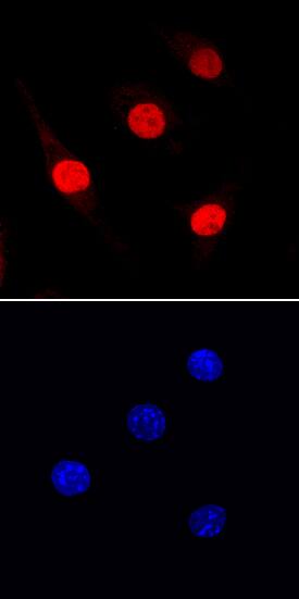
PU.1/Spi-1 was detected in immersion fixed J774A.1 mouse reticulum cell sarcoma macrophage cell line using Rat Anti-Mouse PU.1/Spi-1 Monoclonal Antibody (Catalog # MAB7124) at 10 µg/mL for 3 hours at room temperature. Cells were stained using the NorthernLights™ 557-conjugated Anti-Rat IgG Secondary Antibody (red, upper panel; Catalog # NL013) and counterstained with DAPI (blue, lower panel). Specific staining was localized to nuclei. View our protocol for Fluorescent ICC Staining of Non-adherent Cells.Applications for Mouse PU.1/Spi-1 Antibody
Immunocytochemistry
Sample: Immersion fixed J774A.1 mouse reticulum cell sarcoma macrophage cell line
Western Blot
Sample: NR8383 rat alveolar macrophage cell line and J774A.1 mouse reticulum cell sarcoma macrophage cell line
Formulation, Preparation, and Storage
Purification
Reconstitution
Formulation
Shipping
Stability & Storage
- 12 months from date of receipt, -20 to -70 °C as supplied.
- 1 month, 2 to 8 °C under sterile conditions after reconstitution.
- 6 months, -20 to -70 °C under sterile conditions after reconstitution.
Background: PU.1/Spi-1
Spi-1 (SFFV proviral integration 1 protein; also PU.1 and Sfpi1) is a 37-41 kDa member of the ets family of transcription factors. It is both an RNA and DNA binding protein that is found in hematopoietic cells such as B cells, neutrophils, macrophages and dendritic cells (DC). Spi-1 can act as both a transcriptional activator and repressor. In DC, Spi-1 promotes the expression of CD80, CD86 and CD11b, while in proerythroblasts it blocks GATA-1 induced transcriptional activation. Spi-1 binds to DNA as a monomer and interacts with multiple factors such as Runx-1, IRF8, GATA-1 and c-Jun. Mouse Spi-1 is 272 amino acids (aa) in length. It contains a transactivation region (aa 7-117), a PEST domain (aa 118-164), and a DNA-binding domain (aa 165-266). Phosphorylation on Ser41, 142 and 148 increases activity. There is one alternative start site at Met7. Over aa 1-169, mouse Spi-1 shares 88% and 81% aa identity with rat and human Spi-1, respectively.
Long Name
Alternate Names
Gene Symbol
UniProt
Additional PU.1/Spi-1 Products
Product Documents for Mouse PU.1/Spi-1 Antibody
Product Specific Notices for Mouse PU.1/Spi-1 Antibody
For research use only

