PD-1 Antibody (J43) [PE]
Novus Biologicals, part of Bio-Techne | Catalog # NBP1-43700


Conjugate
Catalog #
Key Product Details
Species Reactivity
Mouse
Applications
Flow Cytometry
Label
PE (Excitation = 488 nm, Emission = 575 nm)
Antibody Source
Monoclonal Armenian Hamster IgG Clone # J43
Concentration
0.2 mg/ml
Product Specifications
Immunogen
The immunogen for this antibody was PD1.
Clonality
Monoclonal
Host
Armenian Hamster
Isotype
IgG
Scientific Data Images for PD-1 Antibody (J43) [PE]
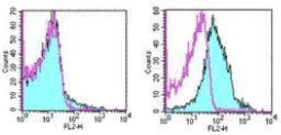
Flow Cytometry: PD-1 Antibody (J43) [PE] [NBP1-43700] - See notes for image caption
Applications for PD-1 Antibody (J43) [PE]
Application
Recommended Usage
Flow Cytometry
1:10-1:1000
Application Notes
The J43 antibody has been tested by flow cytometric analysis of mouse Con-A activated spleen cell suspensions and mouse PD-1 transfected cells. This can be used at less than or equal to 0.5 ug per test. Cell number should be determined empirically but can range from 10^5to 10^8cells/test.
Formulation, Preparation, and Storage
Purification
Protein A or G purified
Formulation
PBS (pH 7.2) with 0.1% gelatin
Preservative
0.09% Sodium Azide
Concentration
0.2 mg/ml
Shipping
The product is shipped with polar packs. Upon receipt, store it immediately at the temperature recommended below.
Stability & Storage
Store at 4C in the dark.
Background: PD-1
Long Name
Programmed Death-1
Alternate Names
CD279, PD1, PDCD1, SLEB2
Gene Symbol
PDCD1
Additional PD-1 Products
Product Documents for PD-1 Antibody (J43) [PE]
Product Specific Notices for PD-1 Antibody (J43) [PE]
This product is for research use only and is not approved for use in humans or in clinical diagnosis. Primary Antibodies are guaranteed for 1 year from date of receipt.
Loading...
Loading...
Loading...
Loading...
Loading...
Loading...