Rat GFR alpha-1/GDNF R alpha-1 Antibody
R&D Systems, part of Bio-Techne | Catalog # AF560


Key Product Details
Validated by
Biological Validation
Species Reactivity
Validated:
Rat
Cited:
Human, Mouse, Rat, Porcine, Avian - Chicken, Transgenic Mouse
Applications
Validated:
Blockade of Receptor-ligand Interaction, Immunohistochemistry, Western Blot
Cited:
Flow Cytometry, IFC, Immunochromatography, Immunocytochemistry, Immunohistochemistry, Immunohistochemistry-Frozen, Immunohistochemistry-Paraffin, Neutralization, Western Blot
Label
Unconjugated
Antibody Source
Polyclonal Goat IgG
Product Specifications
Immunogen
Mouse myeloma cell line NS0-derived recombinant rat GFR alpha‑1/GDNF R alpha‑1
Asp25-Leu445
Accession # Q62997
Asp25-Leu445
Accession # Q62997
Specificity
Detects rat GFR alpha‑1/GDNF R alpha‑1 in direct ELISAs and Western blots. In direct ELISAs, approximately 20% cross-reactivity with recombinant human GFR alpha‑1 is observed and less than 1% cross-reactivity with recombinant mouse GFR alpha‑2 is observed.
Clonality
Polyclonal
Host
Goat
Isotype
IgG
Scientific Data Images for Rat GFR alpha-1/GDNF R alpha-1 Antibody
Detection of Rat GFR alpha‑1/GDNF R alpha‑1 by Western Blot.
Western blot shows lysates of rat brain tissue. PVDF membrane was probed with 0.2 µg/mL of Goat Anti-Rat GFRa-1/GDNF Ra-1 Antigen Affinity-purified Polyclonal Antibody (Catalog # AF560) followed by HRP-conjugated Anti-Goat IgG Secondary Antibody (Catalog # HAF109). A specific band was detected for GFRa-1/GDNF Ra-1 at approximately 52 kDa (as indicated). This experiment was conducted under non-reducing conditions and using Immunoblot Buffer Group 1.GFR alpha‑1/GDNF R alpha‑1 in Rat Spinal Cord.
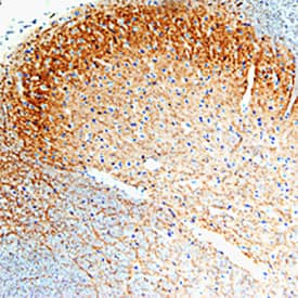
GFRa-1/GDNF Ra-1 was detected in perfusion fixed frozen sections of rat spinal cord using Goat Anti-Rat GFRa-1/GDNF Ra-1 Antigen Affinity-purified Polyclonal Antibody (Catalog # AF560) at 15 µg/mL overnight at 4 °C. Tissue was stained using the Anti-Goat HRP-DAB Cell & Tissue Staining Kit (brown; Catalog # CTS008) and counterstained with hematoxylin (blue). Specific staining was localized to spinal cord dorsal horn. View our protocol for Chromogenic IHC Staining of Frozen Tissue Sections.Detection of Mouse GFR alpha-1/GDNF R alpha-1 by Immunocytochemistry/ Immunofluorescence
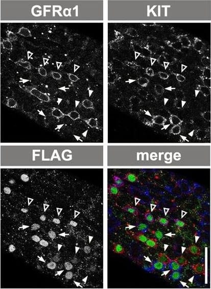
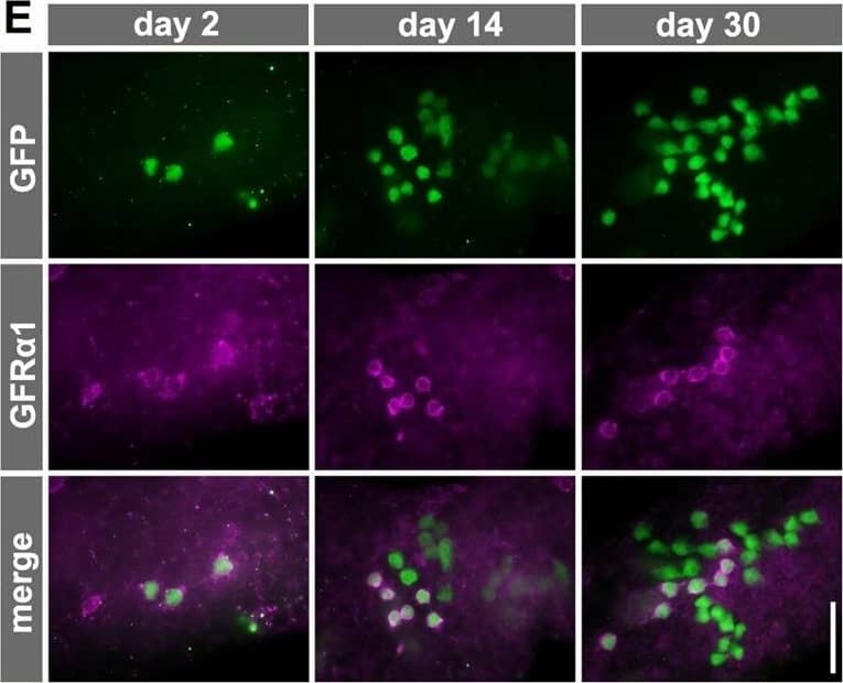
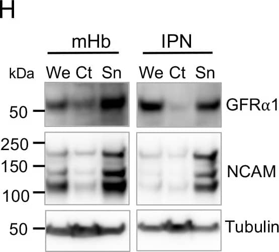
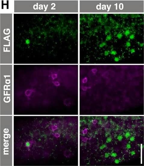
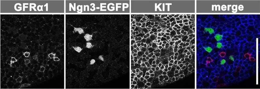
Ectopic RAR gamma expression by GFR alpha1+ spermatogonia. (A) The CAG-CAT-3xFLAG-Rarg transgene. When CAT between the loxP sites is deleted by TM-activated Cre, FLAG-tagged RAR gamma is constitutively expressed under the control of the CAG promoter. (B) Experimental design of the fate analysis of GFR alpha1+ cells with enforced FLAG-RAR gamma expression upon VA readministration in VAD mice, as shown in C-F. Gfra1-CreERT2; CAG-CAT-3xFLAG-Rarg transgenic mice were maintained in VAD and VA was administered 2 days after TM injection, as indicated. Testes were then processed for IF. (C,D) IF images of whole-mount seminiferous tubules of the mice described above, 2 days after VA injection, stained for FLAG-RAR gamma (green) and KIT (magenta) (C), and cell number relative to the number of initial induced cells (D). Data for GFP-labeled NGN3+ and GFR alpha1+ cells are reproduced from Fig. 2C and Fig. 3C, respectively, for comparison. The mean±s.e.m. value of three testes is shown. *P<0.003 (t-test), compared with the values of FLAG-RAR gamma+ GFR alpha1+ cells at day 2. (E,F) Representative confocal images of the same field of whole-mounts of seminiferous tubules of mice treated as described above, at 2 days after VA injection; staining was performed for GFR alpha1, KIT and FLAG (E). Open arrowheads, white arrowheads and small arrows indicate FLAG+ cells that are GFR alpha1+/KIT+, GFR alpha1+/KIT− and GFR alpha1−/KIT+, respectively. (F) Quantitation of GFP+ and FLAG-RAR gamma+ cells showing different patterns of GFR alpha1 and KIT expression in Gfra1-CreERT2; CAG-CAT-EGFP and Gfra1-CreERT2; CAG-CAT-3xFLAG-Rarg mice, respectively. Cell numbers are shown above each bar. (G) Experimental design of the fate analysis of GFR alpha1+ cells with enforced FLAG-RAR gamma expression under normal conditions, as shown in H-J. Gfra1-CreERT2; CAG-CAT-3xFLAG-Rarg transgenic mice were pulsed with TM at 13-17 weeks of age, and after 2 and 10 days their testes were processed for IF. (H) IF images of whole-mount seminiferous tubules 2 and 10 days after TM injection, stained for FLAG-RAR gamma and GFR alpha1. (I,J) Numbers of GFR alpha1+ Aundiff (magenta), GFR alpha1− Aundiff (green), KIT+ (blue) spermatogonia and total cells (black) in either GFP-labeled (I) or FLAG-RAR gamma-expressing (J) cells of Gfra1-CreERT2; CAG-CAT-EGFP and Gfra1-CreERT2; CAG-CAT-3xFLAG-Rarg mice, respectively, following the schedule shown in G. The mean±s.e.m. of four (I) and three (J) testes are shown. *P<0.05 (t-test), compared with the values on day 2. Scale bars: 50 μm. Image collected and cropped by CiteAb from the following open publication (https://journals.biologists.com/dev/article/doi/10.1242/dev.118695/258623/Hierarchical-differentiation-competence-in), licensed under a CC-BY license. Not internally tested by R&D Systems.Applications for Rat GFR alpha-1/GDNF R alpha-1 Antibody
Application
Recommended Usage
Blockade of Receptor-ligand Interaction
Immunohistochemistry
5-15 µg/mL
Sample: Perfusion fixed frozen sections of rat spinal cord
Sample: Perfusion fixed frozen sections of rat spinal cord
Western Blot
0.2 µg/mL
Sample: Rat brain tissue
Sample: Rat brain tissue
Reviewed Applications
Read 14 reviews rated 4.7 using AF560 in the following applications:
Formulation, Preparation, and Storage
Purification
Antigen Affinity-purified
Reconstitution
Reconstitute at 0.2 mg/mL in sterile PBS. For liquid material, refer to CoA for concentration.
Formulation
Lyophilized from a 0.2 μm filtered solution in PBS with Trehalose. *Small pack size (SP) is supplied either lyophilized or as a 0.2 µm filtered solution in PBS.
Shipping
Lyophilized product is shipped at ambient temperature. Liquid small pack size (-SP) is shipped with polar packs. Upon receipt, store immediately at the temperature recommended below.
Stability & Storage
Use a manual defrost freezer and avoid repeated freeze-thaw cycles.
- 12 months from date of receipt, -20 to -70 °C as supplied.
- 1 month, 2 to 8 °C under sterile conditions after reconstitution.
- 6 months, -20 to -70 °C under sterile conditions after reconstitution.
Background: GFR alpha-1/GDNF R alpha-1
‑2 are members of a family of at least four cysteine-rich glycosyl-phosphatidylinositol (GPI)-linked cell surface proteins that share conserved placements of many of their cysteine residues. Binding of GDNF to membrane-associated GFR alpha-1 or GFR alpha-2 initiates the association with and activation of the Ret tyrosine kinase. Soluble GFR alphas released enzymatically from the cell surface-associated protein with phosphatidylinositol phospholipase C, as well as recombinantly produced soluble GFR alpha-1, can also bind with high-affinity to GDNF and trigger the activation of Ret tyrosine kinase. Rat GFR alpha-1 cDNA encodes a 468 amino acid (aa) residue protein with an
N‑terminal 24 aa residue hydrophobic signal peptide. Like other GPI-linked proteins, rat GFR alpha-1 has a C-terminal hydrophobic region which is preceded by a three aa residue (ASS) GPI-binding site. Human GFR alpha-1 shares 93% amino acid identity with rat GFR alpha-1. The expression of the various GFR alphas are differentially regulated in the central and peripheral nervous system, suggesting complementary roles for the GFR alphas in mediating the activities of the GDNF family of neurotrophic factors.
References
- Thompson, J. et al. (1998) Mol. Cell Neurosci. 11:117.
- Trupp, M. et al. (1998) Mol. Cell Neurosci. 11:47.
- Baloh, R.H. et al. (1998) Proc. Natl. Acad. Sci. USA 95:5801.
Long Name
Glial Cell line-derived Neurotrophic Factor Receptor alpha 1
Alternate Names
GDNF R alpha-1, GFR alpha1, GFRa-1, GFRA1
Gene Symbol
GFRA1
UniProt
Additional GFR alpha-1/GDNF R alpha-1 Products
Product Documents for Rat GFR alpha-1/GDNF R alpha-1 Antibody
Product Specific Notices for Rat GFR alpha-1/GDNF R alpha-1 Antibody
For research use only
Loading...
Loading...
Loading...
Loading...
