Human Phospho-Tie-2 DuoSet IC ELISA
R&D Systems, part of Bio-Techne | Catalog # DYC2720-2

Key Product Details
Assay Type
Sample Type
Reactivity
Human Phospho-Tie-2 DuoSet IC ELISA Features
- Optimized capture and detection antibody pairings and recommended concentrations save lengthy development time
- Development protocols are provided to guide further assay optimization
- Assay can be customized to your specific needs
- Available in 2, 5, and 15-(96-well) plate pack sizes
- Economical alternative to Western blot
Product Summary for Human Phospho-Tie-2 DuoSet IC ELISA
Product Specifications
Assay Format
Sample Volume Required
Detection Method
Conjugate
Specificity
Label
Scientific Data Images for Human Phospho-Tie-2 DuoSet IC ELISA
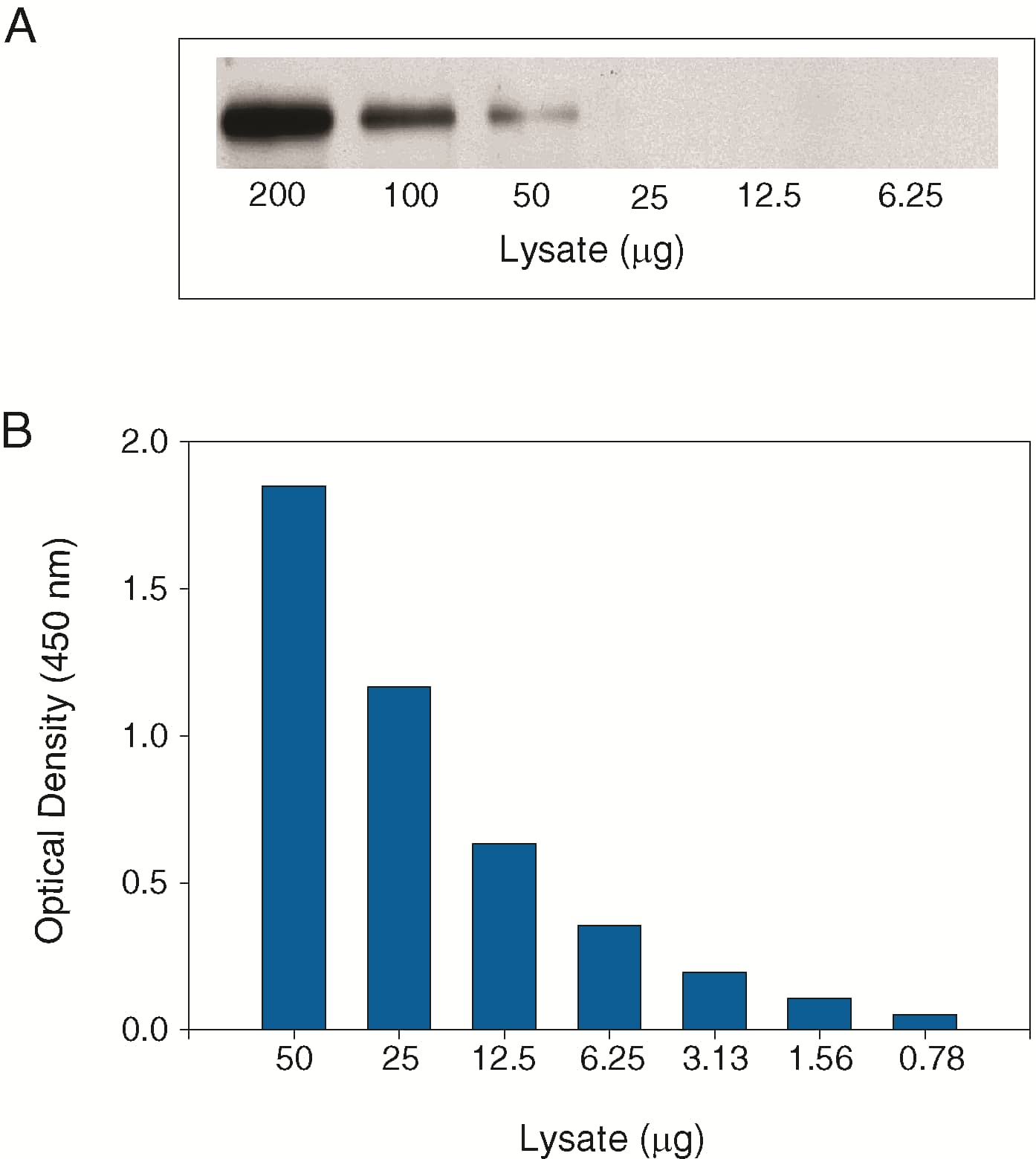
Figure 1. The Human Phospho-Tie-2 DuoSet IC ELISA is more sensitive than immunoprecipitation (IP)-Western analysis
Human Tie-2 transfected Baf/3 cells (Baf/3-hTie-2) were treated with 600 ng/mL recombinant human Ang-1 for seven minutes to induce tyrosine phosphorylation of Tie-2. Serial dilutions of lysates were analyzed by (A) IP-Western blot and (B) this ELISA. IPs were done using an anti-Tie-2 monoclonal antibody and goat anti-mouse agarose. Immunoblots were incubated with a biotinylated anti-phosphotyrosine monoclonal antibody (Catalog # BAM1676) to detect phospho-Tie-2. Bands were visualized with Streptavidin-HRP (Cat # DY998) followed by chemiluminescent detection using WesternGloTM Chemiluminescent Detection Substrate (Catalog # AR004).Figure 2. The Human Phospho-Tie-2 DuoSet IC ELISA detects ligand-induced Tie-2 tyrosine phosphorylation
Baf/3-hTie-2 cells were untreated or treated with 600 ng/mL recombinant human Ang-1 for seven minutes. ELISA and IP-Western blot (inset) analyses were done using 50 μg and 200 μg of lysate, respectively. IP-Western blots for phospho-Tie-2 (p-Tie-2) were done as described in Figure 1. Blots were stripped and total Tie-2 was detected using a biotinylated anti-Tie-2 polyclonal antibody (Catalog # BAF313). Human Phospho-Tie-2 can be detected in this ELISA by using approximately 4 times less lysate than is needed for a conventional IP-Western blot.Figure 3. The specificity of the Human Phospho-Tie-2 DuoSet IC ELISA is confirmed by receptor competition
Baf/3-hTie-2 cells were treated with 600 ng/mL recombinant human Ang-1 for seven minutes. The indicated amounts of recombinant extracellular domains of human Tie-2(Catalog #313-TI), human Tie-1 (Catalog #619-TI), human VEGF R2 (Catalog #357-KD) or human VEGF R3 (Catalog #321-FL) were added to 50 μg lysate and analyzed using this ELISA. Competition was observed only with recombinant Tie-2.Kit Contents for Human Phospho-Tie-2 DuoSet IC ELISA
- Capture Antibody
- Conjugated Detection Antibody
- Calibrated Immunoassay Standard or Control
Other Reagents Required
PBS: (Catalog # DY006), or 137 mM NaCl, 2.7 mM KCl, 8.1 mM Na2HPO4, 1.5 mM KH2O4, pH 7.2 - 7.4, 0.2 µm filtered
Wash Buffer: (Catalog # WA126), or equivalent
Lysis Buffer*
IC Diluent*
Blocking Buffer*
Substrate Solution: 1:1 mixture of Color Reagent A (H2O2) and Color Reagent B (Tetramethylbenzidine) (Catalog # DY999)
Stop Solution: 2 N H2SO4 (Catalog # DY994)
Microplates: R&D Systems (Catalog # DY990), or equivalent
Plate Sealers: ELISA Plate Sealers (Catalog # DY992), or equivalent
*For the Lysis Buffer, IC Diluent, and Blocking BUffer recommended for a specific DuoSet ELISA Development Kit, please see the product
Preparation and Storage
Shipping
Stability & Storage
Background: Tie-2
Long Name
Alternate Names
Entrez Gene IDs
Gene Symbol
Additional Tie-2 Products
Product Documents for Human Phospho-Tie-2 DuoSet IC ELISA
Product Specific Notices for Human Phospho-Tie-2 DuoSet IC ELISA
For research use only


