Recombinant Human Activin A Hyperactive Protein, CF
R&D Systems, part of Bio-Techne | Catalog # BT-ACTAH

Key Product Details
Source
CHO
Accession #
Structure / Form
Disulfide-linked homodimer
Conjugate
Unconjugated
Applications
Bioactivity
Product Specifications
Source
Chinese Hamster Ovary cell line, CHO-derived human Activin A protein
Gly311-Ser426, F368A
Gly311-Ser426, F368A
Purity
>95%, by SDS-PAGE visualized with Silver Staining and quantitative densitometry by Coomassie® Blue Staining.
Endotoxin Level
<0.10 EU per 1 μg of the protein by the LAL method.
N-terminal Sequence Analysis
Gly311
Predicted Molecular Mass
13 kDa
SDS-PAGE
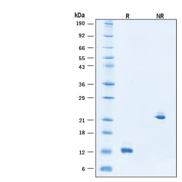
12-14 kDa, under reducing conditions.
Activity
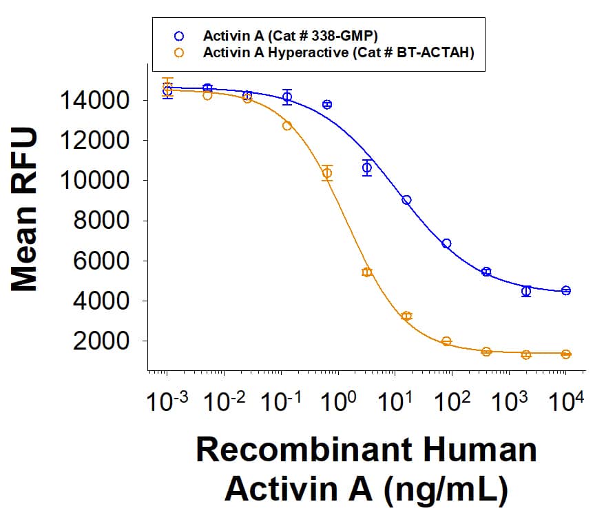
Measured by its ability to induce cytotoxicity using MPC-11 mouse B lymphocyte cells.
The ED50 for this effect is 1.00-15.0 ng/mL.
The ED50 for this effect is 1.00-15.0 ng/mL.
Scientific Data Images for Recombinant Human Activin A Hyperactive Protein, CF
Recombinant Human Activin A Hyperactive Protein Bioactivity.
Human Activin A Hyperactive Protein (Catalog # BT-ACTAH) induces cytotoxicity on MCP-11 cells. The ED50 for this effect is 1.00-15.0 ng/mL. The hyperactive Activin A protein has greater bioactivity than Wild Type Activin A.Recombinant Human Activin A Hyperactive Protein Bioactivity.
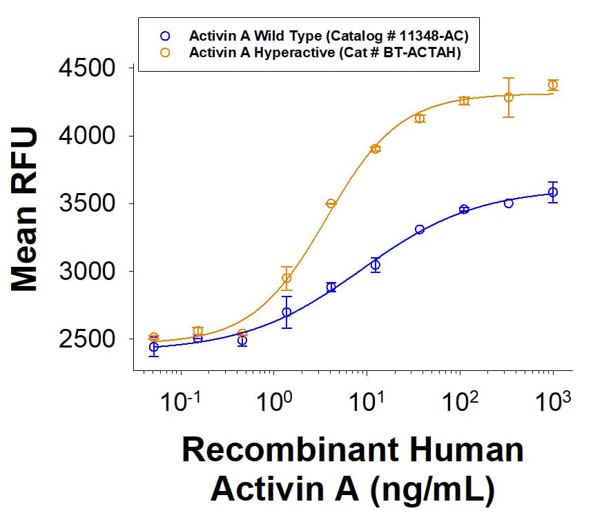
Human Activin A Hyperactive Protein (Catalog # BT-ACTAH) induces SBE (SMAD-binding element) reporter activity in HEK293 human embryonic kidney cells. The hyperactive Activin A protein has greater bioactivity than Wild Type Activin A.Recombinant Human Activin A Hyperactive Protein SDS-PAGE.
2 μg/lane of Recombinant Human Activin A Hyperactive Protein (Catalog # BT-ACTAH) was resolved with SDS-PAGE under reducing (R) and non-reducing (NR) conditions and visualized by Coomassie® Blue staining, showing bands at 12-14 kDa and 20-30 kDa, respectively.Formulation, Preparation and Storage
BT-ACTAH
| Formulation | Lyophilized from a 0.2 μm filtered solution in Acetonitrile and TFA with Trehalose. |
| Reconstitution | Reconstitute the 10 μg size at 100 μg/mL in sterile 4 mM HCl. Reconstitute all other sizes at 500 μg/mL in sterile 4 mM HCl. |
| Shipping | The product is shipped at ambient temperature. Upon receipt, store it immediately at the temperature recommended below. |
| Stability & Storage | Use a manual defrost freezer and avoid repeated freeze-thaw cycles.
|
Background: Activin A
References
- Kumanov, P. et al. (2005) Reprod. Biomed. Online 10:786.
- Maeshima, A. et al. (2008) Endocr. J. 55:1.
- Rodgarkia-Dara, C. et al. (2006) Mutat. Res. 613:123.
- Werner, S. and C. Alzheimer (2006) Cytokine Growth Factor Rev. 17:157.
- Xu, P. and A.K. Hall (2006) Dev. Biol. 299:303.
- Shav-Tal, Y. and D. Zipori (2002) Stem Cells 20:493.
- Chen, Y.G. et al. (2006) Exp. Biol. Med. 231:534.
- Gray, A.M. and A.J. Mason (1990) Science 247:1328.
- Mason, A.J. et al. (1996) Mol. Endocrinol. 10:1055.
- Thompson, T.B. et al. (2004) Mol. Cell. Endocrinol. 225:9.
- Harrison, C.A. et al. (2005) Trends Endocrinol. Metab. 16:73.
- Onichtchouk, D. et al. (1999) Nature 401:480.
- Gray, P.C. et al. (2002) Mol. Cell. Endocrinol. 188:254.
- Kelber, J.A. et al. (2008) J. Biol. Chem. 283:4490.
- Phillips, D.J. et al. (1997) J. Endocrinol. 155:65.
- Schneyer, A. et al. (2003) Endocrinology 144:1671.
- Ghorbani-Dalini, S. et al. (2020) 3 Biotech. 10:215.
- Mennen, R.H. et al. (2022) Reprod Toxicol. 107:44.
- Mishra, S. et al. (2021) Stem Cells. 39:551.
- Tanigawa, S. et al. (2019) Stem Cell Reports 13:322.
- Cangkrama, M. et al. (2020) Trends Mol. Med. 26:1107.
- Ries, A. et al. (2020) Expert Opin. Ther. Targets. 24:985.
- Pinjusic, K. et al. (2022) J. Immunother. Cancer. 10:e004533.
- Dadheech, N. et al. (2020) Stem Cell Res. Ther. 11:327.
Alternate Names
activin AB alpha polypeptide, Activin beta-A chain, erythroid differentiation factor, Erythroid differentiation protein, follicle-stimulating hormone-releasing protein, FSH-releasing protein, inhibin beta A chain, inhibin beta A subunit, Inhibin, beta-1
Gene Symbol
INHBA
UniProt
Additional Activin A Products
Product Documents for Recombinant Human Activin A Hyperactive Protein, CF
Product Specific Notices for Recombinant Human Activin A Hyperactive Protein, CF
For research use only
Loading...
Loading...
Loading...


