Recombinant Human Glypican 3 His Alexa Fluor® 647 Protein Best Seller
R&D Systems, part of Bio-Techne | Catalog # AFR2119

Key Product Details
Source
Accession #
Structure / Form
Labeled with Alexa Fluor® 647
Excitation Wavelength: 650 nm
Emission Wavelength: 668 nm
Conjugate
Applications
Product Specifications
Source
Gln25-His559 with a C-terminal 6-His tag
Purity
Endotoxin Level
N-terminal Sequence Analysis
Predicted Molecular Mass
SDS-PAGE
Activity
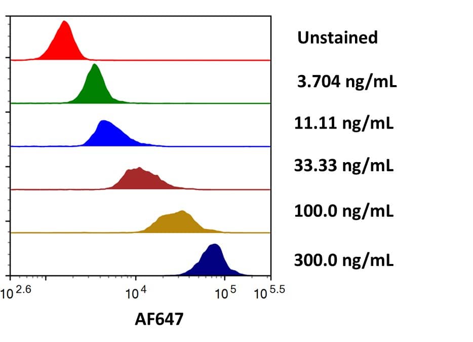
Scientific Data Images for Recombinant Human Glypican 3 His Alexa Fluor® 647 Protein
Flow cytometry analysis for Recombinant Human Glypican 3 His-tag Alexa Fluor® 647 staining on Anti-Human Glypican 3 Antibody conjugated beads.
Streptavidin coated beads conjugated to biotinylated Anti-Human Glypican 3 (BAF2119) were stained with the indicated concentrations of Recombinant Human Glypican 3 His-tag Alexa Fluor® 647 (Catalog # AFR2119).Formulation, Preparation and Storage
AFR2119
| Formulation | Supplied as a 0.2 μm filtered solution in PBS with BSA as a carrier protein. |
| Shipping | The product is shipped with dry ice or equivalent. Upon receipt, store it immediately at the temperature recommended below. |
| Stability & Storage | Protect from light. Use a manual defrost freezer and avoid repeated freeze-thaw cycles.
|
Background: Glypican 3
Glypicans (GPC) are a family of heparan sulfate proteoglycans that are attached to the cell surface by a glycosylphosphatidylinositol (GPI) anchor. Six members of this family have been identified in mammals (GPC1-GPC6). All glypican core proteins contain an N-terminal signal peptide, a large globular cysteine-rich domain (CRD) with 14 invariant cysteine residues, a stalk-like region containing the heparan sulfate attachment sites, and a C-terminal GPI attachment site. While glypican proteins do not share strong amino acid sequence identity (they range from 17-63%), the conserved cysteine residues in their CRDs suggests similarity in their
three‑dimensional structure (1, 2). Mutations in GPC3 cause a rare disorder in humans, Simpson-Golabi-Behmel Syndrome, which is characterized by pre and postnatal overgrowth of multiple tissues and organs and an increased risk for developing embryonic tumors (3). These features are also present in the mouse knock-out of GPC3 indicating that GPC3 regulates cell survival and inhibits cell proliferation during development (4). Glypican 3 has been implicated in regulating many different signaling pathways including: IGF, FGF, BMP and Wnt. An endoproteolytic processing of GPC3 by proprotein convertases is required for the modulation of Wnt signaling (5). Direct interaction with FGF-basic has been observed and is mediated by the heparan sulfate chains (6).
References
- Filmus, J. and S.B. Selleck (2001) J. Clinical Invest. 108:497.
- De Cat, B and G. David (2001) Seminars in Cell & Dev. Biol. 12:117.
- Pilia, G. et al. (1996) Nat. Genet. 12: 241.
- Cano-Gauci, D.F. et al. (1999) J. Cell Biol. 146: 255.
- De Cat, B. et al. (2003) J. Cell Biol. 163:625.
- Song, H.H. et al. (1997) J. Biol. Chem. 272:7574.
Alternate Names
Gene Symbol
UniProt
Additional Glypican 3 Products
Product Documents for Recombinant Human Glypican 3 His Alexa Fluor® 647 Protein
Product Specific Notices for Recombinant Human Glypican 3 His Alexa Fluor® 647 Protein
This product is provided under an agreement between Life Technologies Corporation and R&D Systems, Inc, and the manufacture, use, sale or import of this product is subject to one or more US patents and corresponding non-US equivalents, owned by Life Technologies Corporation and its affiliates. The purchase of this product conveys to the buyer the non-transferable right to use the purchased amount of the product and components of the product only in research conducted by the buyer (whether the buyer is an academic or for-profit entity). The sale of this product is expressly conditioned on the buyer not using the product or its components (1) in manufacturing; (2) to provide a service, information, or data to an unaffiliated third party for payment; (3) for therapeutic, diagnostic or prophylactic purposes; (4) to resell, sell, or otherwise transfer this product or its components to any third party, or for any other commercial purpose. Life Technologies Corporation will not assert a claim against the buyer of the infringement of the above patents based on the manufacture, use or sale of a commercial product developed in research by the buyer in which this product or its components was employed, provided that neither this product nor any of its components was used in the manufacture of such product. For information on purchasing a license to this product for purposes other than research, contact Life Technologies Corporation, Cell Analysis Business Unit, Business Development, 29851 Willow Creek Road, Eugene, OR 97402, Tel: (541) 465-8300. Fax: (541) 335-0354.
This product is provided under an agreement between Life Technologies Corporation and R&D Systems, Inc, and the manufacture, use, sale or import of this product is subject to one or more US patents and corresponding non-US equivalents, owned by Life Technologies Corporation and its affiliates. The purchase of this product conveys to the buyer the non-transferable right to use the purchased amount of the product and components of the product only in research conducted by the buyer (whether the buyer is an academic or for-profit entity). The sale of this product is expressly conditioned on the buyer not using the product or its components (1) in manufacturing; (2) to provide a service, information, or data to an unaffiliated third party for payment; (3) for therapeutic, diagnostic or prophylactic purposes; (4) to resell, sell, or otherwise transfer this product or its components to any third party, or for any other commercial purpose. Life Technologies Corporation will not assert a claim against the buyer of the infringement of the above patents based on the manufacture, use or sale of a commercial product developed in research by the buyer in which this product or its components was employed, provided that neither this product nor any of its components was used in the manufacture of such product. For information on purchasing a license to this product for purposes other than research, contact Life Technologies Corporation, Cell Analysis Business Unit, Business Development, 29851 Willow Creek Road, Eugene, OR 97402, Tel: (541) 465-8300. Fax: (541) 335-0354.
For research use only
