Breadcrumb
- Home
- Products
- Neural Stem Cells
- Neural Stem Cells Stem Cell Products
- Rat Cortical Stem Cells (3 x 10e6 cells/vial) (NSC001)
Rat Cortical Stem Cells (3 x 10e6 cells/vial)
R&D Systems, part of Bio-Techne | Catalog # NSC001

Key Product Details

Summary for Rat Cortical Stem Cells (3 x 10e6 cells/vial)
Kit Summary
For cortical stem cell expansion and differentiation into neurons, astrocytes, and oligodendrocytes.
Key Benefits
- Confirmed multipotency reduces experimental variation
- Highly pure cells that are ready to use
- Each lot confirmed for high Nestin expression
Why use Pre-isolated Fully Characterized Rat Cortical Stem Cells?
Although there are a number of protocols available to isolate rat cortical stem cells, each isolation can yield cells that vary in yield, purity, and their ability to proliferate. See Details
R&D Systems offers Rat Cortical Stem Cells that are of high purity, frozen at a Passage 0, and are validated to be free of mycoplasma and microbial contamination. Rat primary cortical stem cells were isolated from the cortex of embryonic E14.5 Sprague-Dawley rats. The cells were cultured in a monolayer system in medium supplemented with N-2 Plus Media Supplement (Catalog # AR003) or N-2 MAX Media Supplement (Catalog # AR009) and Recombinant Human FGF basic (Catalog # 233-FB or 4114-TC). Cells were then harvested and cryopreserved. The cells are designated as passage 0 (P0) cells.
R&D Systems Rat Cortical Stem Cells:
- Have been fully characterized to reduce experimental variation.
- Have been shown to differentiate into neurons, astrocytes, and oligodendrocytes.
- Can be expanded as monolayers or neurospheres.
- Are free of mycoplasma and microbial contamination.
Components
1 vial of Rat Cortical Stem Cells containing 3 x 10⁶ cells. See Details
Storage
Store in liquid nitrogen for up to 1 year.
Quality Control
- Cells from this lot have been thawed and tested for their ability to proliferate using either a monolayer system (for 3 passages) or a neurosphere system (for 4 passages). Stem and progenitor cells expanded from the end of passage 3 (monolayer system) or passage 4 (neurosphere system) have been examined for Nestin and SOX2 expression. They were also tested for their ability to differentiate into astrocytes, neurons, and oligodendrocytes.
- The cells tested negative for mycoplasma using the MycoProbe™ Mycoplasma Detection Kit (Catalog # CUL001B). The cells also tested negative for microbial contamination.
Precautions
This product contains trace amounts of DMSO and human transferrin. The transferrin was purified from plasma that was tested at the donor level using an FDA licensed method and found to be non-reactive for anti-HIV 1/2 and Hepatitis B surface antigen.
Neural stem cells provide an excellent model for research focused on neural development and neurological disorders. R&D Systems offers ready-to-use primary cortical stem cells isolated from E14.5 Sprague-Dawley rats. In addition, primary mouse cortical stem cells isolated from E14.5 CD-1 mice are available. Every lot of R&D Systems Cortical Stem Cells is validated for a high level of Nestin expression and the capacity for multi-lineage differentiation into astrocytes, neurons, and oligodendrocytes. Our cortical stem cells are tested to ensure highest quality and lot to lot consistency. Both rat and mouse Cortical Stem Cells can be optimally expanded as monolayers or neurospheres.
To complement the use of primary neural stem cells, we offer a range of supportive products, including culture media which is specifically optimized for use with neural stem cells. We offer kits to promote the in vitro proliferation of neural precursors, and kits to differentiate neural stem cells into dopaminergic neurons or oligodendrocytes. In addition, kits are available which contain panels of antibodies designed to monitor the differentiation and identification of neural precursors, astrocytes, neurons, and oligodendrocytes.
| Species | Rat |
| Source | N/A |

|
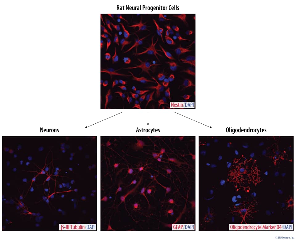
Verification of Neural Progenitor Cell Multipotency Following Expansion with N-2 MAX Media Supplement. Rat Cortical Stem Cells were differentiated for 7 days in media supplemented with N-2 MAX Media Supplement (Catalog # AR009). Differentiated cells were stained with a Mouse Neuron-specific Anti-beta-III Tubulin Monoclonal Antibody (Catalog # MAB1195) followed by the NorthernLights™ (NL)557-conjugated Secondary Antibody (Catalog # NL007; red) to detect neurons. The cells were stained with a Sheep Anti-Human/Rat GFAP Antigen Affinity-purified Polyclonal Antibody (Catalog # AF2594) followed by the NL557-conjugated Secondary Antibody (red; Catalog # NL010) to detect astrocytes; and cells were stained with a Mouse Anti-Human/Mouse/Rat/Chicken Oligodendrocyte Marker O4 Monoclonal Antibody (Catalog # MAB1326), followed by the NL557-conjugated Secondary Antibody (Catalog # NL019) to detect oligodendrocytes. The nuclei were counterstained with DAPI (blue). |

|
Neural Progenitor Cells Expanded with N-2 Plus Media Supplement Express Nestin and SOX2. Rat Cortical Stem Cells (Catalog # NSC001) were cultured for 7 days in media supplemented with 1X N-2 Plus Media Supplement (Catalog # AR003) and 20 ng/mL of Recombinant Human FGF basic (Catalog # 233-FB). The cells were stained with a PE-conjugated Mouse Anti-Human Nestin Monoclonal Antibody (Catalog # IC1259P, red histogram), a PE-conjugated Mouse Anti-Human/Mouse SOX2 Monoclonal Antibody (Catalog # IC2018P, green histogram), or a PE-conjugated Mouse IgG2A Isotype Control Antibody (Catalog # IC003P, open histogram). Under these conditions, cells were shown to express high levels of neural multipotency markers Nestin and SOX2. |

|
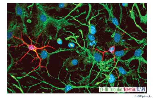
Differentiation of Rat Cortical Stem Cells. Neural progenitors were cultured for seven days in DMEM/F12 containing N-2 Plus Media Supplement (Catalog # AR003) without FGF Basic to induce differentiation. The differentiated cells were labeled with a Goat Anti-Rat Nestin Affinity-purified Polyclonal Antibody (Catalog # AF2736) followed by the NorthernLights™(NL)493-conjugated Donkey Anti-Goat IgG Secondary Antibody (Catalog # NL003; green) and a Mouse Neuron-specific Anti-beta-III Tubulin Monoclonal Antibody (Catalog # MAB1195) followed by the NL-557-conjugated Donkey IgG Anti-Mouse Secondary Antibody (Catalog # NL007; red). Nuclei were counterstained with DAPI (blue). |
Preparation & Storage
| Shipping Conditions | The product is shipped with dry ice or equivalent. Upon receipt, store it immediately at the temperature recommended below. |
| Storage | Store in liquid nitrogen for up to 12 months from the date of receipt. |
Assay Procedure
Refer to the product datasheet for complete product details.
Briefly, Rat Cortical Stem Cells can be thawed and expanded by the neurosphere or monolayer system:
- Thaw Rat Cortical Stem Cells
- Plate cells in Completed NSC Base Media containing FGF basic
- Expand cells using the monolayer or neurosphere system
Reagents & Materials
Reagents Supplied in Rat Cortical Stem Cells (Catalog # NSC001)
- 1 vial of Rat Cortical Stem Cells containing 3 x 10⁶ cells.
Other Supplies Required
Expanding Rat Cortical Stem Cells Using the Neurosphere System
Reagents
- N-2 Plus Media Supplement (Catalog # AR003)
- Recombinant FGF basic (Catalog # 233-FB)
- Bovine Fibronectin (Catalog # 1030-FN)
- PBS
- DMEM/F12
- Glucose
- Glutamine
- NaHCO3
- Penicillin-Streptomycin 100X
- Poly-L-ornithine
- Ca2+/Mg2+-free Hank's Balanced Salt Solution (HBSS) (10X)
- HEPES
- BSA, very low endotoxin
- Trypan Blue, 0.4%
- Deionized (DI) water
Materials
- 10 cm tissue culture plates
- 50 mL centrifuge tubes
- 0.2 µm, sterile filter units
- Plastic cell scraper
- Pipettes and pipette tips
Equipment
- 37 °C and 5% incubator
- Centrifuge
- Hemocytometer
- Microscope
Other Supplies Required
Expanding Rat Cortical Stem Cells Using the Monolayer System
Reagents
- N-2 Plus Media Supplement (Catalog # AR003) or N-2 MAX Media Supplement (Catalog # AR009)
- Recombinant FGF basic (Catalog # 233-FB)
- Bovine Fibronectin (Catalog # 1030-FN)
- PBS
- DMEM/F12
- Glucose
- Glutamine
- NaHCO3
- BSA, very low endotoxin
- Trypan Blue, 0.4%
- Acetic Acid
- Deionized (DI) water
Materials
- 6-well plates
- 15 mL centrifuge tubes
- Pasteur pipettes
- Pipettes and pipette tips
- 0.2 μm, sterile filter unit
Equipment
- 37 °C and 5% incubator
- Centrifuge
- Hemocytometer
- Microscope
Procedure Overview for Expanding Rat Cortical Stem Cells Using the
Neurosphere System
Thawing Cryopreserved Rat Cortical Stem Cells

- Pipette up and down and as cells thaw, transfer the thawed portion into a 50 mL tube containing pre-warmed Completed Base NSC Media supplemented with 20 ng/mL FGF basic.
- Centrifuge the cells at 200 x g for 5 minutes.

- Resuspend the cell pellet in 10 mL of Completed NSC Base Media containing FGF basic.
Neurosphere Expansion

- Perform a cell count.

- Plate cells at approximately 1.0 x 10⁶ NSCs in 5 mL of Completed NSC Base Media supplemented with FGF basic per well in a 6-well plate.
- Incubate the cells at 37 °C and 5% CO2.
- Add fresh FGF basic to the media daily.

- Replace the media every 4 days according to the number of neurospheres present:
- a. Less than 50 neurospheres:
- • Transfer the neurospheres into one well of a 6-well plate using 2.5 mL of fresh media and 2.5 mL of spent/conditioned media.
- b. More than 50 neurospheres:
- • Transfer the media containing the neurospheres to a 15 mL tube.
- • Centrifuge for 5 minutes at 100 x g and remove the media.
- • Resuspend the pellet using a small quantity of fresh Completed NSC Base Media containing FGF basic.
- • Add the neurosphere suspension to 5 mL of fresh Completed Base Media containing FGF basic in one well of a 6-well plate.
- a. Less than 50 neurospheres:
- Passage the cells at 5-6 days or when the neurospheres have a dark clump inside or ruffling on the outside.
Passing Neurosphere

- Transfer the media containing the floating neurospheres to a 15 mL tube.
- Centrifuge for 5 minutes at 100 x g.

- Partially dissociate the neurospheres by pipetting up and down 20 times with a P200 pipette.
- At passage 1 and 2 the cells should be split 1:1. After passage 2 the cells can be split 1:2.
- Add 1 mL of fresh pre-warmed media to the vial of frozen rat cortical stem cells.
- See Details
Procedure Overview for Expanding Rat Cortical Stem Cells Using the
Monolayer System
Thawing Cryopreserved Mouse Cortical Stem Cells

- Add 1 mL of fresh pre-warmed media to the vial of frozen rat cortical stem cells.

- Pipette up and down and as cells thaw.
- Transfer the thawed portion into a 50 mL tube containing pre-warmed Completed Base NSC Media supplemented with 20 ng/mL FGF basic.

- Mix 10 μL of the cell suspension with 10 μL of 0.4% Trypan Blue
- Perform a cell count.
Monolayer Expansion

- Plate 1.0 -1.5 x 10⁶ NSCs in 10 mL of Completed NSC Base Media supplemented with FGF basic onto a Poly-L-ornithine/Fibronectin-coated 10 cm plate.
- Incubate the cells at 37 °C and 5% CO2.

- Replace the media once cells become adherent. After 24 hours, add 10 μL of FGF basic stock (1000X) to the culture.
- Replace the media with fresh Completed NSC Base Media every second day.
- Supplement the media daily with FGF basic.
- Passage the cells when they reach 60-70% confluency.
Passaging Cells

- Wash the cells once with pre-warmed HBSS.
- Add 5 mL of HBSS.
- Incubate at room temperature until the cells round up.

- Scrape the cells from the plate.
- Transfer the cells to a 50 mL centrifuge tube.
- Centrifuge for 5 minutes at 200 x g.
- Remove the supernatant.

- Resuspend the cells in 5 mL of Completed NSC Base Media containing FGF basic.

- Mix 10 μL of the cell suspension with 10 μL of 0.4% Trypan Blue
- Perform a cell count.

- Plate 0.8-1.0 x 10⁶ viable cells in 10 mL of Completed NSC Base Media containing FGF basic onto a Poly-L-ornithine/Fibronectin-coated plate.
- Incubate the cells at 37 °C and 5% CO2.

- Replace the media with fresh Completed NSC Base Media every second day.
- Supplement the media with FGF basic daily.
- Passage the cells after 3 days or when the cells reach 70% confluency.
- Coat cell culture plates with Poly-L-ornithine and Fibronectin.
- See Details

