At Bio-Techne have a broad antibody database covering the area of autophagy - over 1400 reagents in total. Autophagy is the bulk degradation of cytoplasmic components - literally, self-digestion of the cell. Double-membrane vesicles, called autophagosomes, carry unwanted cell components to the lysosomes within an inner autophagic membrane. They then fuse, liberating the autophagic body and its contents into the lumen of the vacuole for degradation. This is a complex process involving at least 16 proteins. LC3, however, is the only one known to form a stable association with the membrane of autophagosomes. It is known to exist in two forms: LC3-I, which is found in the cytoplasm, and LC3-II, which is membrane-bound and is converted from LC3-I to initiate formation and lengthening of the autophagosome. It differs from LC3-I only in the fact that it is covalently modified with lipid extensions (lipidation). Detection of this conversion, using LC3 antibodies, is a useful biomarker to detect autophagy – in fact, it’s the only reliable one.

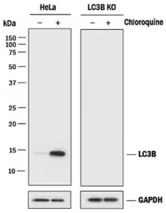
LC3B Knockout Validation: LC3B Antibody [NB100-2220] - Western blot shows lysates of HeLa human cervical epithelial carcinoma parental cell line and LC3B knockout HeLa cell line (KO) untreated (-) or treated (+) with 50 μM Chloroquine for 18 hours. Polyvinylidene difluoride (PVDF) membrane was probed with 0.5 μg/mL of Rabbit Anti-LC3B Monoclonal Antibody [NB100-2220] followed by HRP-conjugated Anti-Rabbit IgG Secondary Antibody [HAF008]. A specific band was detected for LC3B at approximately 15 kDa in the parental HeLa cell line, but is not detectable in the knockout HeLa cell line. GAPDH is shown as a loading control. This experiment was conducted under reducing conditions.
LC3-II is present in both the internal and external compartments of the autophagosome. The association of LC3-II to the nascent phagophore is orchestrated by the activity of the Atg5-Atg12 complex. LC3-II in the forming phagophore interacts with adaptor proteins for the engulfment and eventual processing of cellular components in the autophagolysosome. Overall, because of its principal role in the autophagic process, analysis of LC3 levels via western blot has proved to be an efficient way to monitor autophagic activity. The featured autophagy products in our antibody catalog include wide selections of quality guaranteed LC3, LC3B, Bcl2, ATG5 and HIF-1 alpha reagents. Bio-Techne offers many LC3 reagents for your research needs including: