His Tag Antibody Best Seller
R&D Systems, part of Bio-Techne | Catalog # MAB050


Conjugate
Catalog #
Key Product Details
Species Reactivity
Validated:
Multi-Species
Cited:
Human, Mouse, Rat, Bacteria, Bacteria - Mycobacterium smegmatis, Bacteria - Streptococcus suis, Bovine, Cynomolgus Monkey, E. coli, Guinea Pig, Hamster, Insect, N/A, Primate, Transgenic Bacteria, Transgenic Mouse, Virus, Yeast
Applications
Validated:
CyTOF-ready, Flow Cytometry, Immunoaffinity Purification, Simple Western, Western Blot
Cited:
Affinity Purification, Binding Assay, Bioassay, Co-IP, Cross-linking, ELISA (Capture), ELISA (detection), ELISA Development, ELISA Development (Capture), Epitope Mapping, Flow Cytometry, Functional Assay, Immunocytochemistry, Immunohistochemistry, Immunoprecipitation, Knockdown, Neutralization, Surface Plasmon Resonance, Surface Plasmon Resonance (SPR, Western Blot
Label
Unconjugated
Antibody Source
Monoclonal Mouse IgG1 Clone # AD1.1.10
Product Specifications
Immunogen
His-tagged peptide
Specificity
Detects proteins containing accessible consecutive histidine regions. The antibody detects His tags localized at the amino- or carboxyl-terminus.
Clonality
Monoclonal
Host
Mouse
Isotype
IgG1
Endotoxin Level
<0.20 EU per 1 μg of the antibody by the LAL method.
Scientific Data Images for His Tag Antibody
Detection of His Tag by Western Blot.
Western blot shows lysates of HEK293 human embryonic kidney cell line either non-transfected or transfected with His-tagged human CIQ4 and human MYCOC and CHO Chinese hamster ovary cell line transfected with His-tagged human BAI-1. PVDF membrane was probed with 0.2 µg/mL of Mouse Anti-His Tag Monoclonal Antibody (Catalog # MAB050) followed by HRP-conjugated Anti-Mouse IgG Secondary Antibody (Catalog # HAF018). Specific bands were detected for His Tag at approximately 32, 65, and 120 kDa (as indicated). This experiment was conducted under reducing conditions and using Immunoblot Buffer Group 1.Detection of His Tag in HEK293 Human Cell Line Transfected with His-tagged Proteins by Flow Cytometry.
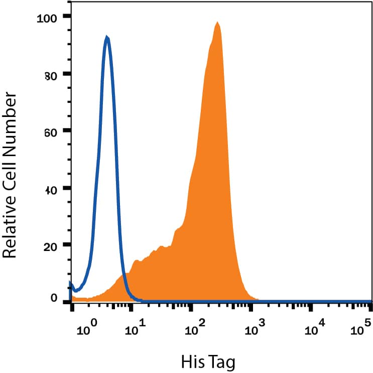
HEK293 human embryonic kidney cell line transfected with His-tagged proteins was stained with Mouse Anti-His Tag Monoclonal Antibody (Catalog # MAB050, filled histogram) or isotype control antibody (Catalog # MAB002, open histogram), followed by Phycoerythrin-conjugated Anti-Mouse IgG Secondary Antibody (Catalog # F0102B). To facilitate intracellular staining, cells were fixed with Flow Cytometry Fixation Buffer (Catalog # FC004) and permeabilized with Flow Cytometry Permeabilization/Wash Buffer I (Catalog # FC005).SARS-Cov-2 Spike 1 His tag protein binding to ACE-2-transfected Human Cell Line is detected by His tag Antibody.
Recombinant SARS-Cov-2 Spike 1 His-tagged protein (10522-CV) binds to HEK293 human embryonic kidney cell line transfected with recombinant human ACE-2 and was detected with Mouse Anti-His Allophycocyanin-conjugated Monoclonal Antibody (IC050A, filled histogram). No staining was observed in the absence of protein (open histogram). Staining was performed using our Staining Membrane-Associated Proteins protocol.Applications for His Tag Antibody
Application
Recommended Usage
CyTOF-ready
Ready to be labeled using established conjugation methods. No BSA or other carrier proteins that could interfere with conjugation.
Flow Cytometry
0.25 µg/106 cells
Sample: HEK293 human embryonic kidney cell line transfected with His-tagged proteins and SARS-Cov-2 Spike 1 His tag protein binding to ACE-2-transfected Human Cell Line
Sample: HEK293 human embryonic kidney cell line transfected with His-tagged proteins and SARS-Cov-2 Spike 1 His tag protein binding to ACE-2-transfected Human Cell Line
Immunoaffinity Purification
Immobilized anti-His antibody can be used to affinity purify proteins containing exposed His tags. The column capacity must be determined for each individual His-tagged protein.
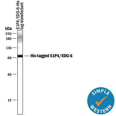
Simple Western
10 µg/mL
Sample: CHO Chinese hamster ovary cell line transfected with His-tagged Human S1P4/EDG-6
Sample: CHO Chinese hamster ovary cell line transfected with His-tagged Human S1P4/EDG-6
Western Blot
0.2 µg/mL
Sample: HEK293 human embryonic kidney cell line and CHO Chinese hamster ovary cell line transfected with His-tagged proteins
Sample: HEK293 human embryonic kidney cell line and CHO Chinese hamster ovary cell line transfected with His-tagged proteins
Reviewed Applications
Read 14 reviews rated 4.8 using MAB050 in the following applications:
Formulation, Preparation, and Storage
Purification
Protein A or G purified from hybridoma culture supernatant
Reconstitution
Reconstitute at 0.5 mg/mL in sterile PBS. For liquid material, refer to CoA for concentration.
Formulation
Lyophilized from a 0.2 μm filtered solution in PBS with Trehalose. *Small pack size (SP) is supplied either lyophilized or as a 0.2 µm filtered solution in PBS.
Shipping
Lyophilized product is shipped at ambient temperature. Liquid small pack size (-SP) is shipped with polar packs. Upon receipt, store immediately at the temperature recommended below.
Stability & Storage
Use a manual defrost freezer and avoid repeated freeze-thaw cycles.
- 12 months from date of receipt, -20 to -70 °C as supplied.
- 1 month, 2 to 8 °C under sterile conditions after reconstitution.
- 6 months, -20 to -70 °C under sterile conditions after reconstitution.
Background: His Tag
Long Name
Histidine Tag
Alternate Names
polyHistidine
Additional His Tag Products
Product Documents for His Tag Antibody
Product Specific Notices for His Tag Antibody
For research use only
Loading...
Loading...
Loading...
Loading...
Loading...
Loading...


