Human CD4 APC-conjugated Antibody
R&D Systems, part of Bio-Techne | Catalog # FAB3791A


Conjugate
Catalog #
Key Product Details
Validated by
Knockout/Knockdown
Species Reactivity
Validated:
Human
Cited:
Human
Applications
Validated:
Flow Cytometry
Cited:
Flow Cytometry
Label
Allophycocyanin (Excitation = 620-650 nm, Emission = 660-670 nm)
Antibody Source
Monoclonal Mouse IgG2A Clone # 11830
Product Specifications
Immunogen
Recombinant Human CD4.
Extracellular Domain
Accession # P01730
Extracellular Domain
Accession # P01730
Specificity
Detects human CD4 in direct ELISAs.
Clonality
Monoclonal
Host
Mouse
Isotype
IgG2A
Scientific Data Images for Human CD4 APC-conjugated Antibody
Detection of CD4 in Human Blood Lymphocytes by Flow Cytometry.
Human peripheral blood lymphocytes were stained with (A) Mouse Anti-Human CD4 APC-conjugated Monoclonal Antibody (Catalog # FAB3791A) or (B) Mouse IgG2AAllophycocyanin Isotype Control (Catalog # IC003A) and Mouse Anti-Human CD3e CFS-conjugated Monoclonal Antibody (Catalog # FAB100F). View our protocol for Staining Membrane-associated Proteins.Detection of Mouse CD4 by Western Blot
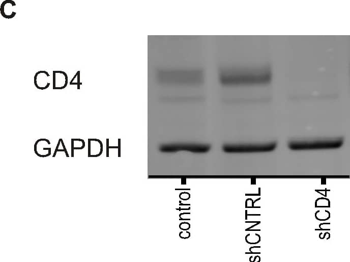
CD4 knock-down in genetically modified stem cell-derived macrophages.A) Schematic diagrams of self-inactivating lentiviral constructs used to express a shRNA (non-specific short-hairpin control (shCNTRL) or a short hairpin targeting CD4 (shCD4)) and puromycin resistance gene (puroR). The Gene Of Interest (GOI) linked to expression of PuromycinR can easily be cloned downstream from the EF1 alpha promoter. B) Detection of CD4 transcripts. RNA was isolated from control and transgenic PSC-macrophages and analysed by RT-qPCR. Symbols represent the relative mean number of copies of CD4 mRNA±SEM of technical replicates (n = 3) using pooled RNA from three independent experiments. C) Detection of protein expression of CD4. Control and transgenic PSC-macrophages lysates were analysed by western blotting using anti-CD4 antibodies. GAPDH, a loading control, was detected using anti-GAPDH antibody. Representative blot is shown.D) Protein levels were measured with Odyssey software (Li-COR) and CD4 expression was normalised to GAPDH expression. Symbols represent mean normalised CD4 expression relative to the PSC-macrophages control group, ±SEM (n = 3 independent experiments). E) Detection of surface protein expression of CD4. Control and transgenic PSC-macrophages were tested for surface CD4 expression by flow cytometry using two different clones of anti-CD4 antibodies. Representative histograms showing CD4 surface staining with mAb clone 11830 (red/brown line, left panel) and with mAb clone OKT4 (blue line, right panel), both compared to isotype control (shaded gray). The expected phenotype (presence or absence of endogenous human CD4) in cells expressing this lentiviral vector is indicated by the symbols. F) Quantification of CD4 expression with mAb clone 11830 (left) and with mAb clone OKT4 (right) relative to the PSC-macrophages control group. The bars reflect the ratio of the geometric mean fluorescence intensity (MFI) over the isotype control ±SEM of independent experiments (n = 4). Image collected and cropped by CiteAb from the following publication (https://dx.plos.org/10.1371/journal.pone.0086071), licensed under a CC-BY license. Not internally tested by R&D Systems.Applications for Human CD4 APC-conjugated Antibody
Application
Recommended Usage
Flow Cytometry
10 µL/106 cells
Sample: Human peripheral blood lymphocytes
Sample: Human peripheral blood lymphocytes
Formulation, Preparation, and Storage
Purification
Protein A or G purified from hybridoma culture supernatant
Formulation
Supplied in a saline solution containing BSA and Sodium Azide.
Shipping
The product is shipped with polar packs. Upon receipt, store it immediately at the temperature recommended below.
Stability & Storage
Protect from light. Do not freeze.
- 12 months from date of receipt, 2 to 8 °C as supplied.
Background: CD4
Alternate Names
CD4
Entrez Gene IDs
Gene Symbol
CD4
UniProt
Additional CD4 Products
Product Documents for Human CD4 APC-conjugated Antibody
Product Specific Notices for Human CD4 APC-conjugated Antibody
For research use only
Loading...
Loading...
Loading...
Loading...
Loading...
Loading...
