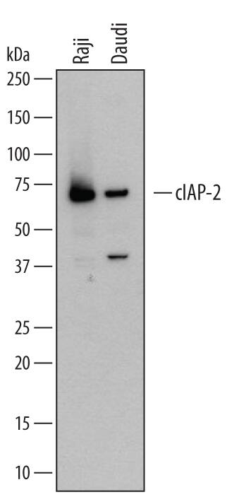
Detection of Human cIAP-2/HIAP-1 by Western Blot
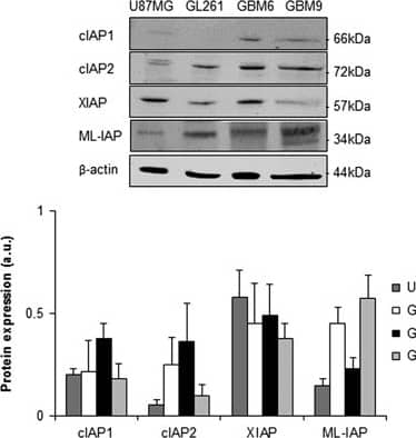
IAP expression in glioblastoma cell lines. Expression levels of cIAP1, cIAP2, XIAP and ML-IAP were analyzed by western blotting and quantified in U87MG and GL261 adherent GBM cell lines, and in GBM6 and GBM9 spheres. Expression level of beta-actin served as loading control. The four GBM cell lines expressed heterogeneously cIAP1, cIAP2, XIAP and ML-IAP. A representative experiment of four experiments is shown. Quantification was performed using ImageJ software (National Institutes of Health, Bethesda, MD, USA) and data presented were normalized to beta-actin expression Image collected and cropped by CiteAb from the following publication (https://pubmed.ncbi.nlm.nih.gov/27490930), licensed under a CC-BY license. Not internally tested by R&D Systems.
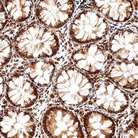
Detection of Human cIAP-2/HIAP-1 by Immunocytochemistry/Immunofluorescence
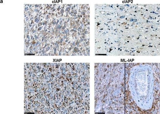
Prognostic value of cIAP1, cIAP2, XIAP and ML-IAP protein expression in human glioblastomas (cohorts 1 and 2). (a) cIAP1-, cIAP2-, XIAP- and ML-IAP-positive stainings in GBM. IAPs were heterogeneously expressed by tumor cells in GBM samples (Table 1). Stainings were diffused with a stronger punctuated positivity into the cytoplasm. Black arrows highlight cIAP2-positive nuclei. Scale bars, 50 μm. (b) Correlation of ML-IAP protein expression with PFS and OS in cohort 1. The cutoff was 35% and was determined by performing a ROC curve. ML-IAP expression of ⩾35% was correlated with a poor prognosis. (c) Correlation of ML-IAP protein expression with PFS and OS in cohort 2. The cutoff was the same as that for cohort 1 analysis (35%). ML-IAP expression of ⩾35% was correlated with a poor prognosis Image collected and cropped by CiteAb from the following publication (https://pubmed.ncbi.nlm.nih.gov/27490930), licensed under a CC-BY license. Not internally tested by R&D Systems.
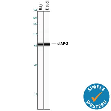
Detection of Human cIAP-2/HIAP-1 by Western Blot
Apoptosis and IAP expression upon SMAC mimetic GDC-0152 treatment in glioblastoma cell lines. (a) Apoptosis (SubG0/G1) of DMSO control and GDC-0152-treated cells was determined by flow cytometry of propidium iodide-stained nuclei and percentage of apoptosis is shown. U87MG and GL261 cell lines were treated for 72 h and GBM6 and GBM9 cell lines were treated for 8 days at the indicated concentrations. At these respective time points, percentage of U87MG cells dead by apoptosis, percentage of GL261 cells, percentage of GBM6 cells and percentage of GBM9 cells. Data are expressed as mean+S.E.M. Three independent experiments were performed for the GL261 cell lines and five for the U87MG, GBM6 and GBM9 cell lines. *P<0.05; **P<0.01; ***P<0.005. (b) Expression levels of cIAP1, cIAP2, XIAP and ML-IAP were analyzed by western blotting. Cell lines were treated with 1 μM of GDC-0152. U87MG and GL261 were treated for 72 h and GBM6 and GBM9 cell lines for 8 days. In all GBM cell lines GDC-0152 decreased IAP expression. Expression level of beta-actin served as loading control. A representative experiment of three experiments is shown Image collected and cropped by CiteAb from the following publication (https://pubmed.ncbi.nlm.nih.gov/27490930), licensed under a CC-BY license. Not internally tested by R&D Systems.
Detection of Human cIAP-2/HIAP-1 by Western Blot
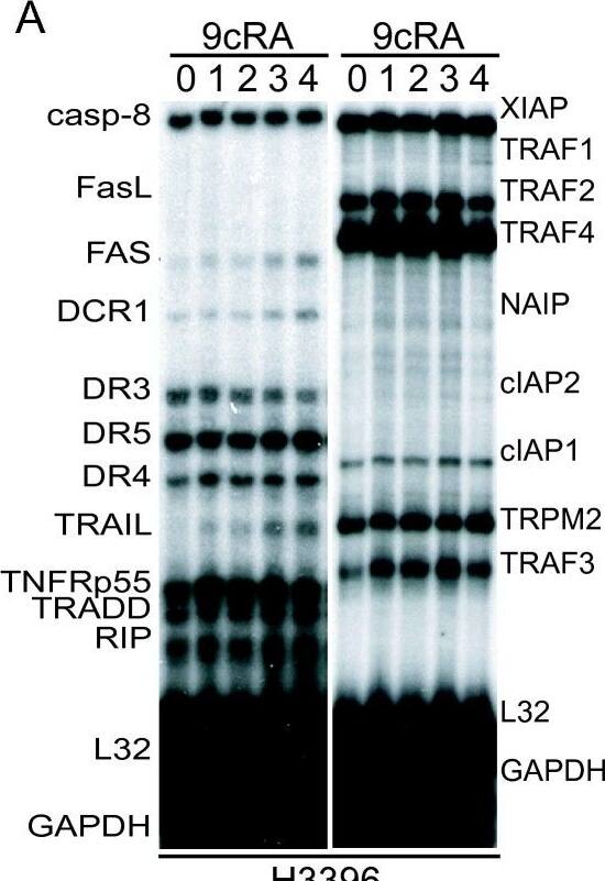
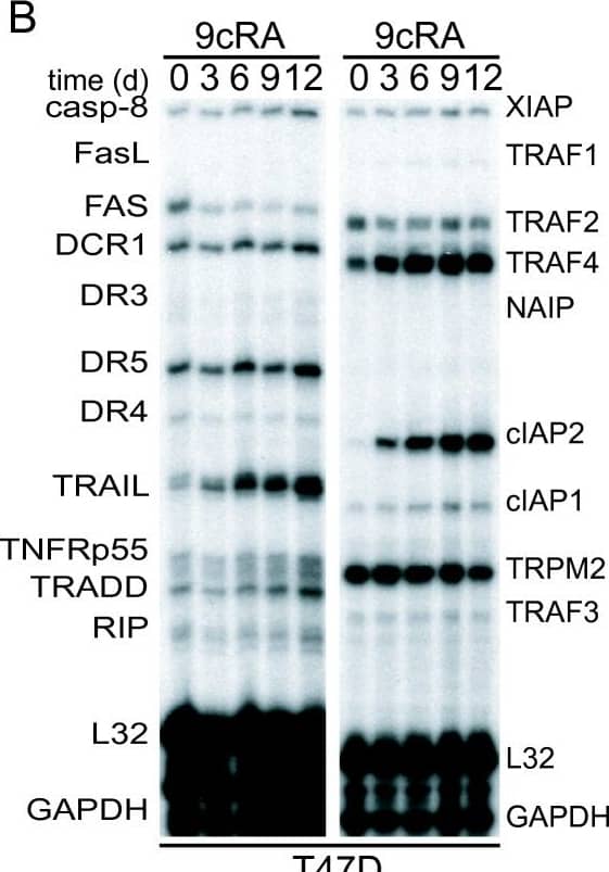
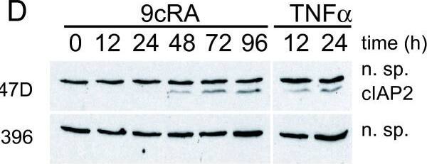
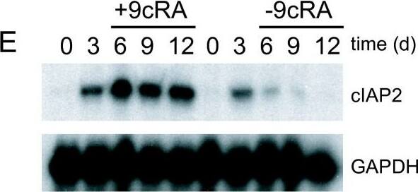
9-cis-RA induces the expression of cIAP2 in breast cancer cells in a cell context dependent manner. (A, B, C) Multiplex RNase protections assays (RPAs) to monitor the effect of 9-cis-RA on the expression of death receptor, death ligands, IAP and TRAF family members in four different breast cancer cell lines. Breast cancer cells were treated for the indicated time with 9-cis-RA at a concentration of 10-6 M. (D) Western blot of whole cell extracts of 9-cis-RA-treated T47D cells and H3396 cells for 0, 12, 24, 48, 72 and 96 hours with anti-cIAP2. The nonspecific signal (n. sp.) confirms equal loading. As a positive control, breast cancer cells were treated with 50 μg/ml of hTNF alpha for 24 and 48 hours. (E) Reversibility of 9-cis-RA-induced cIAP2 gene expression. T47D cells were treated either in the absence or presence of 1 μM 9- cis-RA and after 3 days, total RNA was extracted. In parallel flasks, the medium was removed, and cells were washed and treated with either fresh control medium or medium with 10-6 M of 9-cis-RA and grown for additional 3, 6 and 9 days. Media and ligands were renewed every 3 days. RNA was isolated and analyzed by RPA as described in (A). Equal loading was confirmed by GAPDH RNA level. The images shown are from one representative experiment performed twice with similar results. Image collected and cropped by CiteAb from the following open publication (http://molecular-cancer.biomedcentral.com/articles/10.1186/1476-4598-9-15), licensed under a CC-BY license. Not internally tested by R&D Systems.
Detection of Human cIAP-2/HIAP-1 by Western Blot
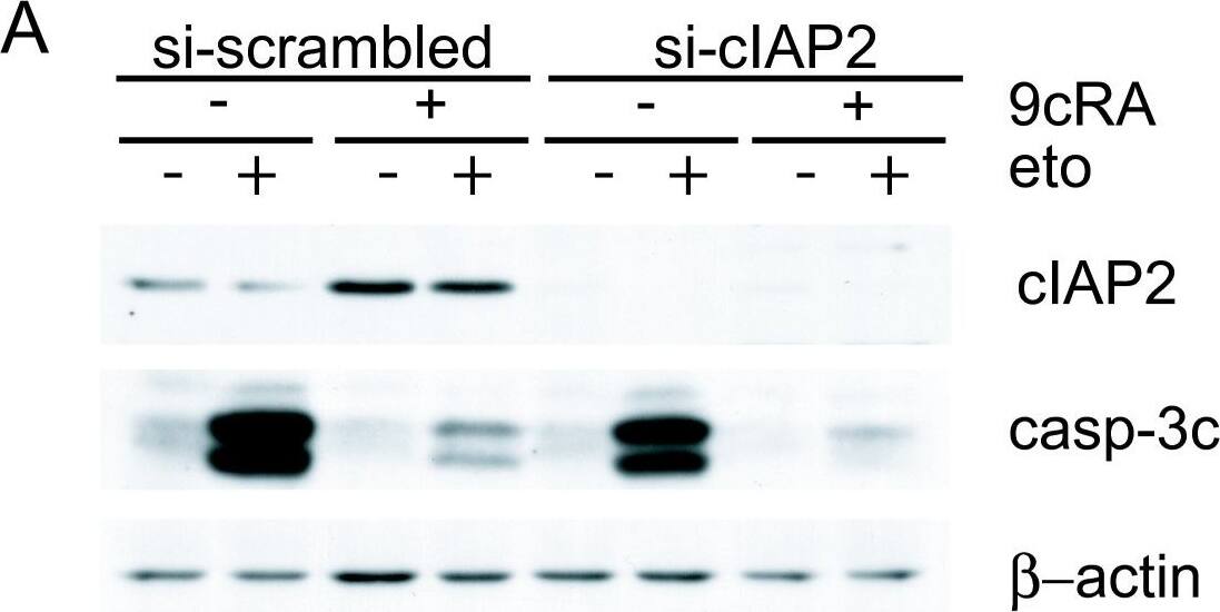
Suppression of cIAP2 expression is not sufficient to abrogate 9-cis-RA inhibition of etoposide-induced apoptosis in T47D cells. (A) T47D cells were transfected with either scrambled-siRNA or cIAP2-siRNA and pretreated with or without 9-cis-RA for 30 h, followed by treatment with etoposide 100 μM for 24 h. Cell lysates were analyzed by western blot for the expression of cleaved caspase-3, cIAP2 and beta-actin using specific antibodies. The images shown are from one representative experiment performed three times with similar results. (B) T47D cells were transfected with either scrambled-siRNA (white bars) or cIAP2-siRNA (black bars). After 24 h, lipid-siRNA complexes were removed from media and cells were pretreated with or without 1 μM 9-cis-RA for 30 h, followed by treatment with etoposide 100 μM for 72 h. The percentage of apoptotic cells was determined by FACS analysis after staining with propidium iodide. The values represent the mean ± SD of three experiments performed in duplicate. Asterisks denote statistically significant differences against the corresponding untreated cells. Image collected and cropped by CiteAb from the following open publication (http://molecular-cancer.biomedcentral.com/articles/10.1186/1476-4598-9-15), licensed under a CC-BY license. Not internally tested by R&D Systems.
Detection of Human cIAP-2/HIAP-1 by Western Blot
9-cis-RA induces the expression of cIAP2 in breast cancer cells in a cell context dependent manner. (A, B, C) Multiplex RNase protections assays (RPAs) to monitor the effect of 9-cis-RA on the expression of death receptor, death ligands, IAP and TRAF family members in four different breast cancer cell lines. Breast cancer cells were treated for the indicated time with 9-cis-RA at a concentration of 10-6 M. (D) Western blot of whole cell extracts of 9-cis-RA-treated T47D cells and H3396 cells for 0, 12, 24, 48, 72 and 96 hours with anti-cIAP2. The nonspecific signal (n. sp.) confirms equal loading. As a positive control, breast cancer cells were treated with 50 μg/ml of hTNF alpha for 24 and 48 hours. (E) Reversibility of 9-cis-RA-induced cIAP2 gene expression. T47D cells were treated either in the absence or presence of 1 μM 9- cis-RA and after 3 days, total RNA was extracted. In parallel flasks, the medium was removed, and cells were washed and treated with either fresh control medium or medium with 10-6 M of 9-cis-RA and grown for additional 3, 6 and 9 days. Media and ligands were renewed every 3 days. RNA was isolated and analyzed by RPA as described in (A). Equal loading was confirmed by GAPDH RNA level. The images shown are from one representative experiment performed twice with similar results. Image collected and cropped by CiteAb from the following open publication (http://molecular-cancer.biomedcentral.com/articles/10.1186/1476-4598-9-15), licensed under a CC-BY license. Not internally tested by R&D Systems.
Detection of Human cIAP-2/HIAP-1 by Western Blot
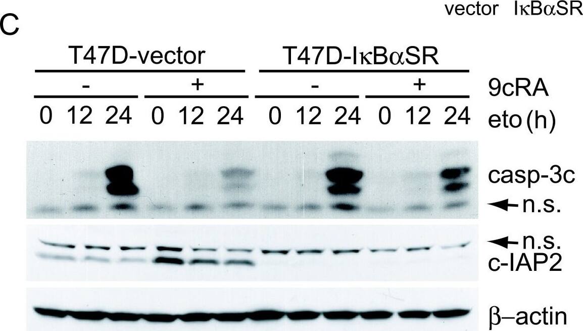
Over-expression of the super-repressor of NF-kappa B activation, I kappaB alpha-SR(S32A/S36A), leads to significant abrogation of retinoic acid-mediated inhibition of etoposide-induced apoptosis. (A) T47D-vector and T47D-I kappaB alphaSR cells were untreated or incubated for the indicated time with hTNF alpha (50 ng/ml). Extracts were analyzed by western blotting with an I kappaB alpha antibody. Equal loading was confirmed with an anti-beta -actin antibody. The images shown are from one representative experiment performed three times with similar results. (B) T47D-vector or T47D-I kappaB alphaSR cells were untreated or treated with 9-cis-RA for 48 h and expression of cIAP2 and beta-actin were analysed by Reverse-Transcriptase Polymerase Reaction and real time PCR. The values represent the mean ± SD of three different experiments performed in duplicate. (C) T47D-vector or T47D-I kappaB alphaSR cells were pretreated with or without 9-cis-RA for 30 h, followed by treatment with etoposide 100 μM as previously indicated. At the indicated times, cell lysates were analyzed by western blot for the expression of cleaved caspase-3, cIAP2 and beta-actin using specific antibodies. The images shown are from one representative experiment performed three times with similar results. (D) T47D-vector or T47D-I kappaB alphaSR cells were pretreated with or without 9-cis-RA for 30 h, followed by treatment with etoposide 100 μM for 72 h. The percentage of apoptotic cells was determined by FACS analysis after staining with propidium iodide. The values represent the mean ± SD of three independent experiments performed in duplicate. Asterisks denote the existence of statistically significant differences between the indicated groups; N.S.: not significant (Student's t-test). Image collected and cropped by CiteAb from the following open publication (http://molecular-cancer.biomedcentral.com/articles/10.1186/1476-4598-9-15), licensed under a CC-BY license. Not internally tested by R&D Systems.

