Human Cyr61/CCN1 Antibody
R&D Systems, part of Bio-Techne | Catalog # AF6009

Key Product Details
Species Reactivity
Applications
Label
Antibody Source
Product Specifications
Immunogen
Ala22-Asp381
Accession # O00622
Specificity
Clonality
Host
Isotype
Scientific Data Images for Human Cyr61/CCN1 Antibody
Detection of Human Cyr61/CCN1 by Western Blot.
Western blot shows lysates of A549 human lung carcinoma cell line and SK-BR-3 human breast cancer cell line. PVDF Membrane was probed with 1 µg/mL of Sheep Anti-Human Cyr61/CCN1 Antigen Affinity-purified Polyclonal Antibody (Catalog # AF6009) followed by HRP-conjugated Anti-Sheep IgG Secondary Antibody (Catalog # HAF016). A specific band was detected for Cyr61/CCN1 at approximately 40 kDa (as indicated). This experiment was conducted under reducing conditions and using Immunoblot Buffer Group 2.Detection of Human Cyr61/CCN1 by Simple WesternTM.
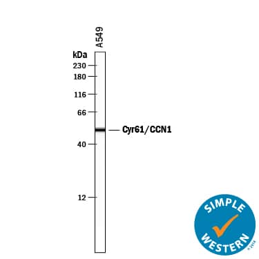
Simple Western lane view shows lysates of A549 human lung carcinoma cell line, loaded at 0.2 mg/mL. A specific band was detected for Cyr61/CCN1 at approximately 52 kDa (as indicated) using 10 µg/mL of Sheep Anti-Human Cyr61/CCN1 Antigen Affinity-purified Polyclonal Antibody (Catalog # AF6009) followed by 1:50 dilution of HRP-conjugated Anti-Sheep IgG Secondary Antibody (Catalog # HAF016). This experiment was conducted under reducing conditions and using the 12-230 kDa separation system.Human Cyr61/CCN1 ELISA Standard Curve.
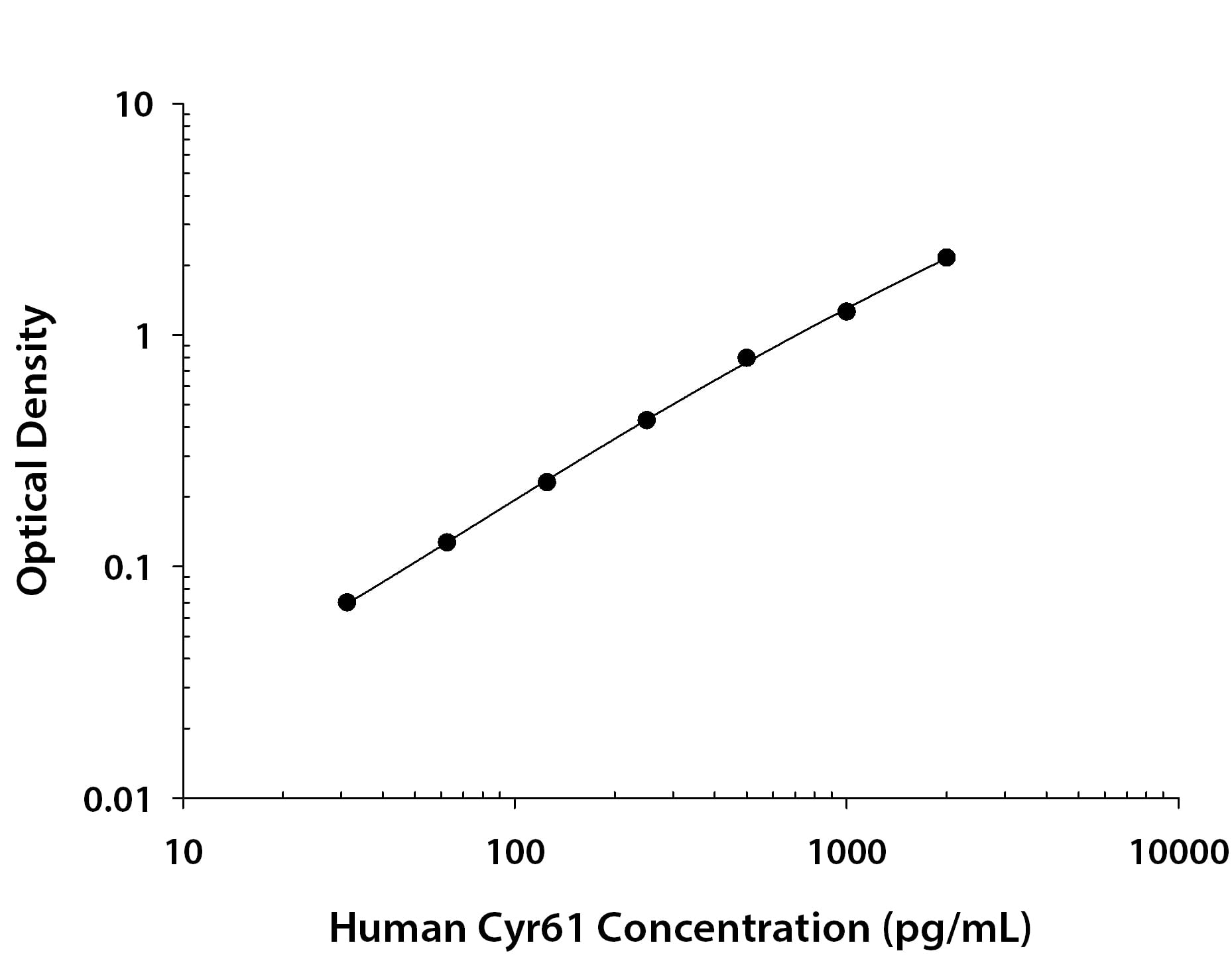
Recombinant Human Cyr61/CCN1 protein was serially diluted 2-fold and captured by Rat Anti-Human Cyr61/CCN1 Monoclonal Antibody (Catalog # MAB40551) coated on a Clear Polystyrene Microplate (Catalog # DY990). Sheep Anti-Human Cyr61/CCN1 Antigen Affinity-purified Polyclonal Antibody (Catalog # AF6009) was biotinylated and incubated with the protein captured on the plate. Detection of the standard curve was achieved by incubating Streptavidin-HRP (Catalog # DY998) followed by Substrate Solution (Catalog # DY999) and stopping the enzymatic reaction with Stop Solution (Catalog # DY994).Applications for Human Cyr61/CCN1 Antibody
ELISA
This antibody functions as an ELISA detection antibody when paired with Rat Anti-Human Cyr61/CCN1 Monoclonal Antibody (Catalog # MAB40551).
This product is intended for assay development on various assay platforms requiring antibody pairs. We recommend the Human Cyr61/CCN1 DuoSet ELISA Kit (Catalog # DY4055) for convenient development of a sandwich ELISA or the Human Cyr61/CCN1 Quantikine ELISA Kit (Catalog # DCYR10) for a complete optimized ELISA.
Simple Western
Sample: A549 human lung carcinoma cell line
Western Blot
Sample: A549 human lung carcinoma cell line and SK‑BR‑3 human breast cancer cell line
Formulation, Preparation, and Storage
Purification
Reconstitution
Formulation
Shipping
Stability & Storage
- 12 months from date of receipt, -20 to -70 °C as supplied.
- 1 month, 2 to 8 °C under sterile conditions after reconstitution.
- 6 months, -20 to -70 °C under sterile conditions after reconstitution.
Background: Cyr61/CCN1
Cysteine-rich angiogenic inducer 61 (Cyr61), also known as CCN1, is a 40‑45 kDa matricellular glycoprotein that plays an important role in cellular adhesion and migration (1). Cyr61 consists of an IGFBP domain, a VWF type C domain, a TSP type I domain, and a cysteine knot domain (2). Mature human Cyr61 shares 93% amino acid sequence identity with mouse and rat Cyr61. It is widely expressed during development and in adult tissues (2, 3). Cyr61 associates with the extracellular matrix (ECM) and with many cell surface molecules including Integrins alphaV beta3, alphaV beta5, alphaM beta2, and alpha6 beta1, Syndecan-4, and heparan sulfate proteoglycans (1, 3). Cyr61 mediates the adhesion and migration of multiple cell types and also promotes vascular endothelial cell tubule formation (4‑6). Plasmin cleavage of
ECM‑bound Cyr61 releases a 28 kDa N-terminal fragment which retains the ability to promote endothelial cell migration (7). Cyr61 exhibits both tumorigenic and tumor suppressor properties. It is upregulated and promotes tumorigenesis, angiogenesis, and metastasis in breast, renal, gastric, squamous cell, and colorectal carcinomas as well as in glioma (8‑12). In contrast, when downregulated, it suppresses tumor growth in endometrial, hepatic, and non-small cell lung cancers (8, 13, 14). Cyr61 is also upregulated in injured skin and bone where it induces the expression of growth factors, cytokines, proteases, and integrins involved in wound repair (15, 16).
References
- Chen, Y. and X.Y. Du (2007) J. Cell. Biochem. 100:1337.
- Jay, P. et al. (1997) Oncogene 14:1753.
- Feng, P. et al. (2008) Int. J. Biochem. Cell Biol. 40:98.
- Chen, N. et al. (2000) J. Biol. Chem. 275:24953.
- Leu, S-J. et al. (2002) J. Biol. Chem. 277:46248.
- Schutze, N. et al. (2007) BMC Cell Biol. 8:45.
- Pendurthi, U.R. et al. (2005) Cancer Res. 65:9705.
- O'Kelly, J. et al. (2008) Int. J. Oncol. 33:59.
- Chintalapudi, M.R. et al. (2008) Carcinogenesis 29:696.
- Xie, D. et al. (2004) Cancer Res. 64:1987.
- Lin, M-T. et al. (2007) J. Biol. Chem. 282:34594.
- Monnier, Y. et al. (2008) Cancer Res. 68:7323.
- Tong, X. et al. (2001) J. Biol. Chem. 276:47709.
- Chien, W. et al. (2004) J. Biol. Chem. 279:53087.
- Hadjiargyrou, M. et al. (2000) J. Bone Miner. Res.15:1014.
- Chen, C-C. et al. (2001) J. Biol. Chem. 276:47329.
Long Name
Alternate Names
Gene Symbol
UniProt
Additional Cyr61/CCN1 Products
Product Documents for Human Cyr61/CCN1 Antibody
Product Specific Notices for Human Cyr61/CCN1 Antibody
For research use only


