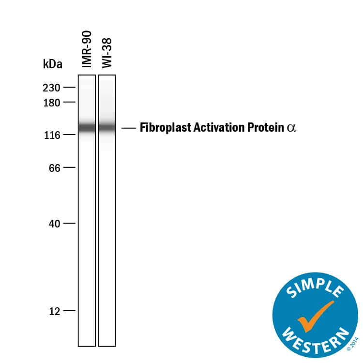
Detection of Human Fibroblast Activation Protein alpha/FAP by Simple WesternTM.
Simple Western lane view shows lysates of IMR-90 human lung fibroblast cell line and WI-38 human lung fibroblast cell line, loaded at 0.2 mg/mL. A specific band was detected for Fibroblast Activation Protein alpha/FAP at approximately 130 kDa (as indicated) using 10 µg/mL of Sheep Anti-Human Fibroblast Activation Protein alpha/FAP Antigen Affinity-purified Polyclonal Antibody (Catalog # AF3715) followed by 1:50 dilution of HRP-conjugated Anti-Sheep IgG Secondary Antibody (
HAF016). This experiment was conducted under reducing conditions and using the 12-230 kDa separation system.
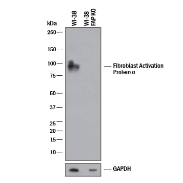
Western Blot Shows Human Fibroblast Activation Protein alpha/FAP Specificity by Using Knockout Cell Line.
Western blot shows lysates of WI-38 human lung fibroblast cell line and human FAP knockout WI-38 human lung fibroblast cell line. PVDF membrane was probed with 0.5 µg/mL of Sheep Anti-Human Fibroblast Activation Protein alpha/FAP Antigen Affinity-purified Polyclonal Antibody (Catalog # AF3715) followed by HRP-conjugated Anti-Sheep IgG Secondary Antibody (
HAF016). A specific band was detected for Fibroblast Activation Protein alpha/FAP at approximately 97 kDa (as indicated) in the parental WI-38 human lung fibroblast cell line, but is not detectable in knockout WI-38 human lung fibroblast cell line. GAPDH (
AF5718) is shown as a loading control. This experiment was conducted under reducing conditions and using Western Blot Buffer Group 1.
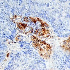
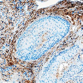
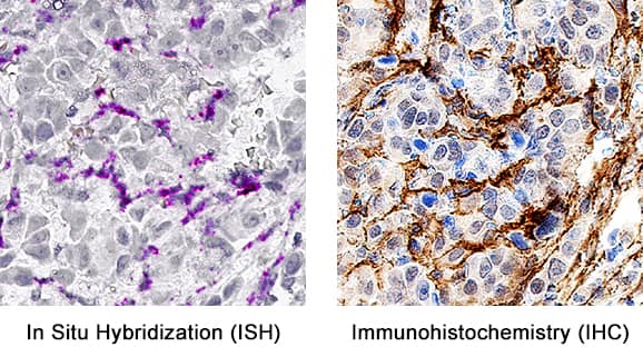
Detection of Fibroblast Activation Protein alpha/FAP in Human Esophagus.
Formalin-fixed paraffin-embedded tissue sections of human squamous cell carcinoma in the esophagus were probed for FAP mRNA (ACD RNAScope Probe, catalog # 411971; Fast Red chromogen, ACD catalog # 322360). Adjacent tissue section was processed for immunohistochemistry using sheep anti-human FAP polyclonal antibody (R&D Systems catalog #
AF3715) at 5ug/mL with overnight incubation at 4 degrees Celsius followed by incubation with anti-sheep IgG VisUCyte HRP Polymer Antibody (Catalog #
VC006) and DAB chromogen (yellow-brown). Tissue was counterstained with hematoxylin (blue). Specific staining was localized to connective tissue.
Detection of Fibroblast Activation Protein alpha /FAP by Western Blot
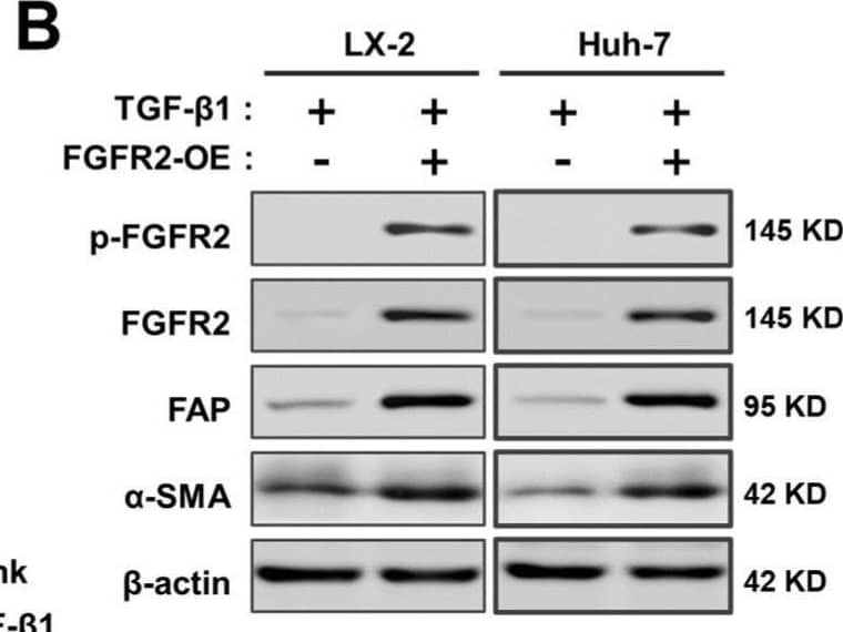
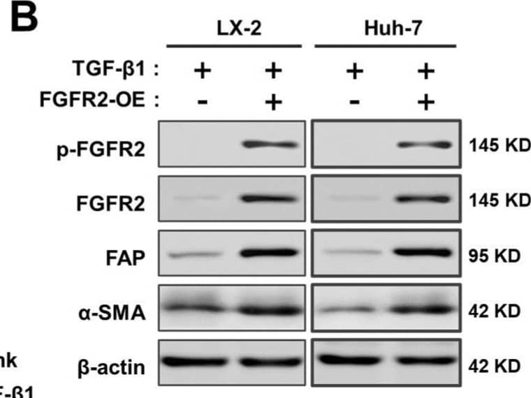
FGFR2 drives the process of liver fibrosis. (A) The expression of liver fibrosis markers (ACTA2, fibroblast activation protein alpha (FAP), alpha-1 type I collagen (COL1A1)) was measured by qPCR. The samples were divided into four groups according to whether cells were induced with or without TGF-beta and whether cells were overexpressed by FGFR2. (B) The expression of markers in wild-type and FGFR2-OE cell lines under TGF-beta induction was analyzed by Western blot analysis. (C) The expression levels of alpha-SMA and (D) collagen secretion in LX-2 and Huh-7 were evaluated, when FGFR2 was overexpressed or knocked down upon equal TGF-beta induction. (E) Cells were collected after 48 h of co-culture, and the expression of alpha-SMA and (F) type I collagen in the lower layer cells was determined by ELISA. The results are marked as significant “*” when p < 0.05, “**” when p < 0.01, and not significant (ns) if p ≥ 0.05. Image collected and cropped by CiteAb from the following open publication (https://pubmed.ncbi.nlm.nih.gov/37111305), licensed under a CC-BY license. Not internally tested by R&D Systems.
Detection of Fibroblast Activation Protein alpha /FAP by Western Blot
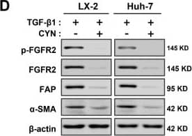
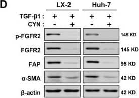
CYN blocks the activation and development of liver fibrosis in vitro. (A) Evaluation of the inhibitory effect of CYN on the fibrosis-promoting effect of FGFR2. The fibrotic transformation of wild-type and FGFR2-OE LX-2 cells and Huh-7 cells was induced through TGF-beta activation, followed by an intervention with CYN for the relevant groups. The expression changes of the fibrosis markers ACTA2 and COL1A1 were analyzed by qPCR. (B) Wild-type cell lines were employed in the aforementioned experiments, and the activation of FGFR2 was triggered by supplementation with the exogenous basic fibroblast growth factor (bFGF) factor. (C) Analysis of the extent of antagonism of CYN towards TGF-beta Signaling. Activation induction models were established by adding or not adding TGF-beta to the cell culture environment with or without the CYN intervention, and the expression of liver fibrosis markers was determined by qPCR, (D) Western blot, and (E,F) ELISA analyses. (G) A co-culture model was used to evaluate the blocking effect of CYN on liver fibrosis activation of signaling transmission. The activation intensity of lower-layer wild-type cells was measured and compared using alpha-SMA expression and collagen secretion. The results are marked as significant “*” when p < 0.05, “**” when p < 0.01, and not significant (ns) if p ≥ 0.05. Image collected and cropped by CiteAb from the following open publication (https://pubmed.ncbi.nlm.nih.gov/37111305), licensed under a CC-BY license. Not internally tested by R&D Systems.
Detection of Fibroblast Activation Protein alpha /FAP by Western Blot
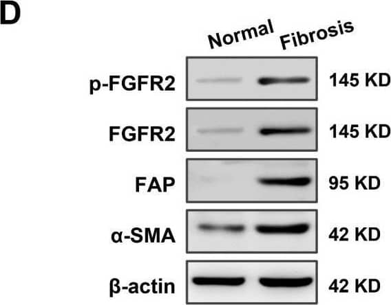
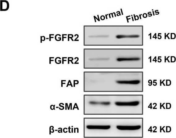
High expression of fibroblast growth factor receptor 2 (FGFR2) coincides with liver fibrosis. (A) The expression of FGFR2 in fibrotic liver and normal liver tissues was evaluated through data mining of human samples affected by Hepatitis B infection (GSE38941), alcohol abuse (GSE28619), and nonalcoholic steatohepatitis (GSE48452), as well as mouse samples induced with carbon tetrachloride (CCl4) (GSE152329, GSE98577). The differential fold expression was determined by comparing the expression of FGFR2 in the fibrotic liver and normal liver tissues. (B) A comparison of changes in FGFR2 expression in liver fibrosis patients in remission (improved) or not in remission (not improved) with mining data of GSE175448. (C) The correlation between FGFR2 expression and the liver fibrosis score (Ishak score) was analyzed by comparing FGFR2 expression in three pairs of liver fibrotic tissues and normal liver tissues through immunohistochemistry. (D) FGFR2 expression in liver fibrosis tissues and normal liver tissues was verified by Western blot and (E) q-PCR analyses. (E) Liver tissues from mice treated with CCl4 for different durations were used to determine the expression of the liver fibrosis markers Actin Alpha 2 (ACTA2) and FGFR2 by q-PCR and the trends of their expression with increasing days of induction. (F) The expression of ACTA2 and (G) FGFR2 was determined by q-PCR in liver tissues from mice treated with CCl4 for varying durations. The statistical significance was determined based on the p-value. The results are marked as significant “*” when p < 0.05, “**” when p < 0.01, “***” when p < 0.001, and not significant (ns) if p ≥ 0.05. Image collected and cropped by CiteAb from the following open publication (https://pubmed.ncbi.nlm.nih.gov/37111305), licensed under a CC-BY license. Not internally tested by R&D Systems.
Detection of Fibroblast Activation Protein alpha /FAP by Western Blot
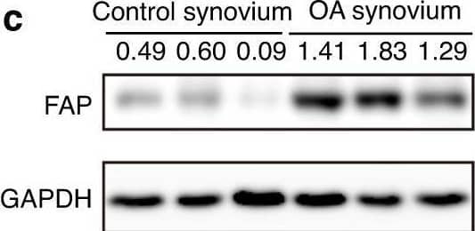
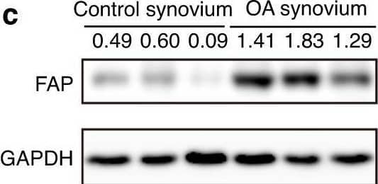
Expression analysis of Fap in OA synovium. a Immunostaining of human FAP in the synovium of control and OA patients. DAPI staining indicates the nucleus. Scale bars: 100 μm. b qPCR analysis of human FAP mRNA levels in the synovium of control and OA patients (n = 8 samples per group). c Western blot analysis of human FAP protein levels in the synovium of control and OA patients (n = 3 samples per group). d Immunostaining of mouse Fap in the knee joint of sham and DMM-treated mice. Sham or DMM surgery was performed in 8-week-old FapLacZ/+ mice, which were sacrificed 8 weeks later, and LacZ immunostaining was used to detect Fap expression at the posterior horn of the medial meniscus (F: femur; T: tibia; M: meniscus; S: synovium). DAPI staining indicates the nucleus. Scale bars: 100 μm. The statistical significance was assessed using two-tailed Student’s unpaired t tests. Data are presented as the mean ± SD (**P < 0.01) Image collected and cropped by CiteAb from the following open publication (https://pubmed.ncbi.nlm.nih.gov/36588124), licensed under a CC-BY license. Not internally tested by R&D Systems.
Detection of Fibroblast Activation Protein alpha /FAP by Western Blot
FGFR2 drives the process of liver fibrosis. (A) The expression of liver fibrosis markers (ACTA2, fibroblast activation protein alpha (FAP), alpha-1 type I collagen (COL1A1)) was measured by qPCR. The samples were divided into four groups according to whether cells were induced with or without TGF-beta and whether cells were overexpressed by FGFR2. (B) The expression of markers in wild-type and FGFR2-OE cell lines under TGF-beta induction was analyzed by Western blot analysis. (C) The expression levels of alpha-SMA and (D) collagen secretion in LX-2 and Huh-7 were evaluated, when FGFR2 was overexpressed or knocked down upon equal TGF-beta induction. (E) Cells were collected after 48 h of co-culture, and the expression of alpha-SMA and (F) type I collagen in the lower layer cells was determined by ELISA. The results are marked as significant “*” when p < 0.05, “**” when p < 0.01, and not significant (ns) if p ≥ 0.05. Image collected and cropped by CiteAb from the following open publication (https://pubmed.ncbi.nlm.nih.gov/37111305), licensed under a CC-BY license. Not internally tested by R&D Systems.
Detection of Fibroblast Activation Protein alpha /FAP by Western Blot
Expression analysis of Fap in OA synovium. a Immunostaining of human FAP in the synovium of control and OA patients. DAPI staining indicates the nucleus. Scale bars: 100 μm. b qPCR analysis of human FAP mRNA levels in the synovium of control and OA patients (n = 8 samples per group). c Western blot analysis of human FAP protein levels in the synovium of control and OA patients (n = 3 samples per group). d Immunostaining of mouse Fap in the knee joint of sham and DMM-treated mice. Sham or DMM surgery was performed in 8-week-old FapLacZ/+ mice, which were sacrificed 8 weeks later, and LacZ immunostaining was used to detect Fap expression at the posterior horn of the medial meniscus (F: femur; T: tibia; M: meniscus; S: synovium). DAPI staining indicates the nucleus. Scale bars: 100 μm. The statistical significance was assessed using two-tailed Student’s unpaired t tests. Data are presented as the mean ± SD (**P < 0.01) Image collected and cropped by CiteAb from the following open publication (https://pubmed.ncbi.nlm.nih.gov/36588124), licensed under a CC-BY license. Not internally tested by R&D Systems.
Detection of Fibroblast Activation Protein alpha /FAP by Western Blot
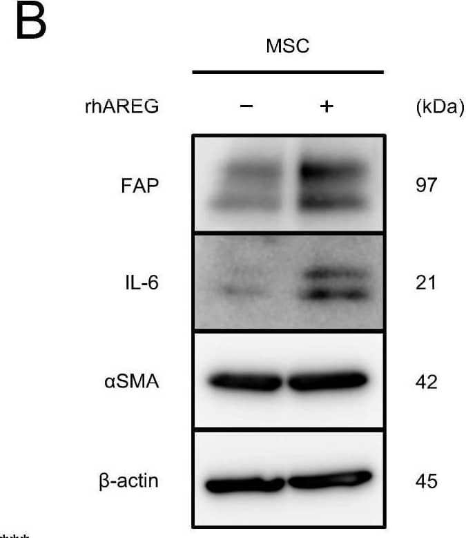
AREG promotes migration and CAF-like differentiation of MSCs: (A,B) To investigate the differentiation of MSCs into CAFs upon treatment with rhAREG (100 ng/mL), the mRNA and protein expression levels of FAP, IL-6, and alphaSMA were compared using qRT-PCR (A) and Western blotting (B). beta-actin was used as a loading control (B). (C,D) The effects of rhAREG (100 ng/mL) on the survival (C) and growth (D) of MSCs were evaluated using the MTS assay. (E) The effect of rhAREG (100 ng/mL) on the migration of MSCs was evaluated using the transwell migration assay. MSCs were seeded in the upper chamber, and migrated cells were counted in five fields of view after 48 h. Representative images for each condition are presented below the graph (E). The data are presented as the mean ± SEM of three independent experiments (A,C–E). N.S., not significant; ** p < 0.01, *** p < 0.001. Scale bars: 100 μm (E). Image collected and cropped by CiteAb from the following open publication (https://pubmed.ncbi.nlm.nih.gov/39451251), licensed under a CC-BY license. Not internally tested by R&D Systems.


