Human HLA-DR Antibody
R&D Systems, part of Bio-Techne | Catalog # MAB11555

Key Product Details
Species Reactivity
Applications
Label
Antibody Source
Product Specifications
Immunogen
Accession # P01903
Specificity
Clonality
Host
Isotype
Scientific Data Images for Human HLA-DR Antibody
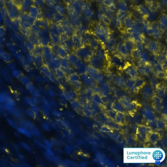
Detection of HLA-DRA in Human Tonsil via Multiplex Immunofluorescence staining on COMET™
HLA-DRA was detected in immersion fixed paraffin-embedded sections of human tonsil using Mouse Anti-Human HLA-DRA Monoclonal Antibody (Catalog # MAB11555) at 1 µg/mL at 37 ° Celsius for 2 minutes. Before incubation with the primary antibody, tissue underwent an all-in-one dewaxing and antigen retrieval preprocessing using PreTreatment Module (PT Module) and Dewax and HIER Buffer H (pH 9). Tissue was stained using the Alexa Fluor™ 555 Goat anti-Mouse IgG Secondary Antibody at 1:100 at 37 ° Celsius for 2 minutes. (Yellow; Lunaphore Catalog # DR555MS) and counterstained with DAPI (blue; Lunaphore Catalog # DR100). Specific staining was localized to the membrane. Protocol available in COMET™ Panel Builder.Detection of Human HLA-DR by Western Blot.
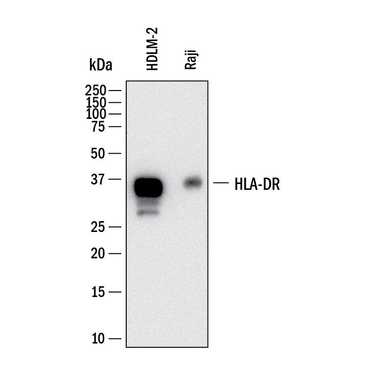
Western Blot shows lysates of HDLM-2 human Hodgkin’s lymphoma cell line and Raji human Burkitt's lymphoma cell line. PVDF membrane was probed with 2 µg/ml of Mouse Anti-Human HLA-DR Monoclonal Antibody (Catalog # MAB11555) followed by HRP-conjugated Anti-Mouse IgG Secondary Antibody (Catalog # HAF018). A specific band was detected for HLA-DR at approximately 35 kDa (as indicated). This experiment was conducted under reducing conditions and using Western Blot Buffer Group 1.Detection of HLA-DR in Human Tonsil.
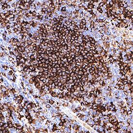
HLA-DR was detected in immersion fixed paraffin-embedded sections of human tonsil using Mouse Anti-Human HLA-DR Monoclonal Antibody (Catalog # MAB11555) at 1 µg/ml for 1 hour at room temperature followed by incubation with the Anti-Mouse IgG VisUCyte™ HRP Polymer Antibody (Catalog # VC001) or the HRP-conjugated Anti-Mouse IgG Secondary Antibody (Catalog # HAF007). Before incubation with the primary antibody, tissue was subjected to heat-induced epitope retrieval using VisUCyte Antigen Retrieval Reagent-Basic (Catalog # VCTS021). Tissue was stained using DAB (brown) and counterstained with hematoxylin (blue). Specific staining was localized to the membrane. View our protocol for Chromogenic IHC Staining of Paraffin-embedded Tissue Sections.Applications for Human HLA-DR Antibody
Immunohistochemistry
Sample: Immersion fixed paraffin-embedded sections of human tonsil
Multiplex Immunofluorescence
Sample: Immersion fixed paraffin-embedded sections of Human Tonsil tissue
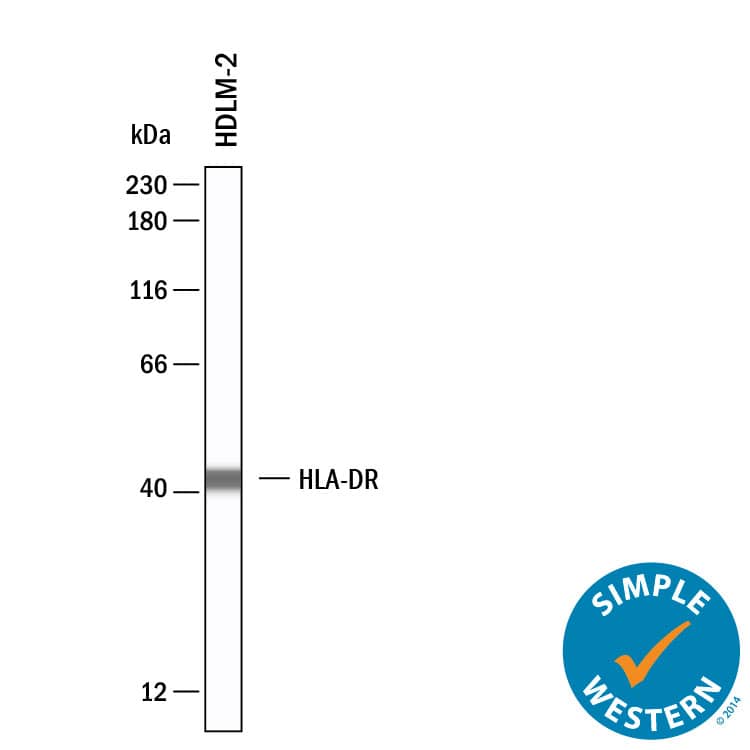
Simple Western
Sample: HDLM-2 human Hodgkin's lymphoma cell line
Western Blot
Sample: HDLM-2 human Hodgkin's lymphoma cell line and Raji human Burkitt's lymphoma cell line
Formulation, Preparation, and Storage
Purification
Reconstitution
Formulation
Shipping
Stability & Storage
- 12 months from date of receipt, -20 to -70 °C as supplied.
- 1 month, 2 to 8 °C under sterile conditions after reconstitution.
- 6 months, -20 to -70 °C under sterile conditions after reconstitution.
Background: HLA-DR
HLA-DR is a transmembrane human major histocompatibility complex 2 (MHC II) family member and consists of a 34 kDa (alpha) subunit and one of several 28 kDa (beta) subunits. HLA-DR is expressed primarily by B cells and dendritic cells (DC), in which it binds peptides derived from internalized and processed antigenic proteins. It presents these peptides on the cell surface for recognition by the T cell receptor on CD4+ T cells. This interaction is central to antigen specificity in the adaptive immune response. HLA-DR alleles, polymorphisms, and aberrant expression are linked to a variety of diseases including autoimmunity and cancer.
Long Name
Alternate Names
Entrez Gene IDs
Gene Symbol
UniProt
Additional HLA-DR Products
Product Documents for Human HLA-DR Antibody
Product Specific Notices for Human HLA-DR Antibody
For research use only


