Human/Mouse/Rat Phospho-FRS2 (Y436) Antibody
R&D Systems, part of Bio-Techne | Catalog # AF5126

Key Product Details
Validated by
Species Reactivity
Validated:
Cited:
Applications
Validated:
Cited:
Label
Antibody Source
Product Specifications
Immunogen
Specificity
Clonality
Host
Isotype
Scientific Data Images for Human/Mouse/Rat Phospho-FRS2 (Y436) Antibody
Detection of Human and Rat Phospho-FRS2 (Y436) by Western Blot.
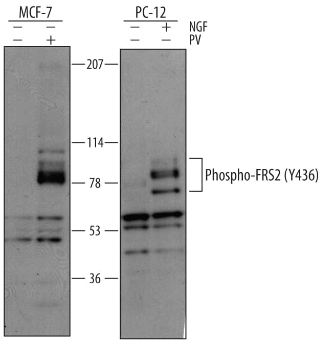
Western blot shows lysates of MCF-7 human breast cancer cell line untreated (-) or treated (+) with 100 µM pervanadate (PV) for 10 minutes and PC-12 rat adrenal pheochromocytoma cell line untreated or treated with 100 ng/mL Recombinant Rat beta-NGF (Catalog # 556-NG) for 10 minutes. PVDF membrane was probed with 1 µg/mL of Rabbit Anti-Human/Mouse/Rat Phospho-FRS2 (Y436) Antigen Affinity-purified Polyclonal Antibody (Catalog # AF5126), followed by HRP-conjugated Anti-Rabbit IgG Secondary Antibody (Catalog # HAF008). Bands were detected for Phospho-FRS2 (Y436) at approximately 70 - 90 kDa (as indicated). This experiment was conducted under reducing conditions and using Immunoblot Buffer Group 1.Phospho-FRS2 (Y436) in A431 Human Cell Line.
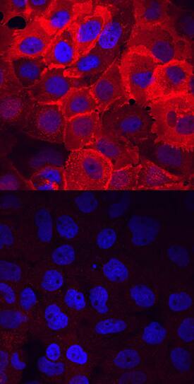
FRS2 phosphorylated at Y436 was detected in immersion fixed A431 human epithelial carcinoma cell line untreated (lower panel) or treated (upper panel) with pervanadate using Rabbit Anti-Human/Mouse/Rat Phospho-FRS2 (Y436) Antigen Affinity-purified Polyclonal Antibody (Catalog # AF5126) at 10 µg/mL for 3 hours at room temperature. Cells were stained using the NorthernLights™ 557-conjugated Anti-Rabbit IgG Secondary Antibody (red; Catalog # NL004) and counterstained with DAPI (blue). View our protocol for Fluorescent ICC Staining of Cells on Coverslips.Applications for Human/Mouse/Rat Phospho-FRS2 (Y436) Antibody
Immunocytochemistry
Sample: Immersion fixed A431 human epithelial carcinoma cell line treated with pervanadate
Western Blot
Sample: Pervanadate-treated MCF-7 human breast cancer cell line and PC-12 rat adrenal pheochromocytoma cell line treated with Recombinant Rat beta-NGF (Catalog # 556-NG)
Formulation, Preparation, and Storage
Purification
Reconstitution
Formulation
Shipping
Stability & Storage
- 12 months from date of receipt, -20 to -70 °C as supplied.
- 1 month, 2 to 8 °C under sterile conditions after reconstitution.
- 6 months, -20 to -70 °C under sterile conditions after reconstitution.
Background: FRS2
FRS2 (FGF R substrate 2; also known as SNT and FRS2-alpha) is a 70‑90 kDa member of the FRS family of lipid-anchored docking proteins. It is an intermediary between FGF and RTK receptors and their Ras/MAPK signaling cascades. FRS2 contains a membrane-anchoring myristoylation signal (aa 1‑6), a PTB domain that interacts with FGF and NGF receptors (aa 13‑115), and a C-terminal tyrosine-rich region that serves as a docking site for GRB-2 and SHP-2 (aa 196‑471). Phosphorylation of Y436 by activated RTKs is required for efficient SHP-2 recruitment.
Long Name
Alternate Names
Gene Symbol
Additional FRS2 Products
Product Documents for Human/Mouse/Rat Phospho-FRS2 (Y436) Antibody
Product Specific Notices for Human/Mouse/Rat Phospho-FRS2 (Y436) Antibody
For research use only

