Human/Mouse TIMP-2 Antibody
R&D Systems, part of Bio-Techne | Catalog # AF971


Key Product Details
Validated by
Species Reactivity
Validated:
Cited:
Applications
Validated:
Cited:
Label
Antibody Source
Product Specifications
Immunogen
Cys27-Pro220
Accession # P16035
Specificity
Clonality
Host
Isotype
Endotoxin Level
Scientific Data Images for Human/Mouse TIMP-2 Antibody
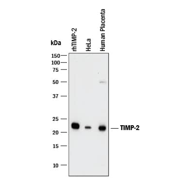
Detection of Human TIMP‑2 by Western Blot.
Western blot shows Recombinant Human TIMP-2 Western Blot Standard Protein (Catalog # WBC023) and lysates of HeLa human cervical epithelial carcinoma cell line and human placenta tissue. PVDF membrane was probed with 1 µg/mL of Goat Anti-Human/Mouse TIMP-2 Antigen Affinity-purified Polyclonal Antibody (Catalog # AF971) followed by HRP-conjugated Anti-Goat IgG Secondary Antibody (Catalog # HAF017). A specific band was detected for TIMP-2 at approximately 22 kDa (as indicated). This experiment was conducted under reducing conditions and using Immunoblot Buffer Group 1.TIMP-2 in Human Ovarian Cancer Tissue.
TIMP-2 was detected in immersion fixed paraffin-embedded sections of human ovarian cancer tissue using 15 µg/mL Goat Anti-Human/Mouse TIMP-2 Antigen Affinity-purified Polyclonal Antibody (Catalog # AF971) overnight at 4 °C. Tissue was stained with the Anti-Goat HRP-DAB Cell & Tissue Staining Kit (brown; Catalog # CTS008) and counter-stained with hematoxylin (blue). View our protocol for Chromogenic IHC Staining of Paraffin-embedded Tissue Sections.Western Blot Shows Human TIMP-2 Specificity by Using Knockout Cell Line.
Western blot shows lysates of HeLa human cervical epithelial carcinoma cell line and human TIMP-2 knockout HeLa human cervical epithelial carcinoma cell line (KO). PVDF membrane was probed with 1 µg/mL of Goat Anti-Human/Mouse TIMP-2 Antigen Affinity-purified Polyclonal Antibody (Catalog # AF971) followed by HRP-conjugated Anti-Goat IgG Secondary Antibody (HAF017). A specific band was detected for TIMP-2 at approximately 22 kDa (as indicated) in the parental HeLa human cervical epithelial carcinoma cell line, but is not detectable in knockout HeLa human cervical epithelial carcinoma cell line. GAPDH (AF5718) is shown as a loading control. This experiment was conducted under reducing conditions and using Western Blot Buffer Group 1.Applications for Human/Mouse TIMP-2 Antibody
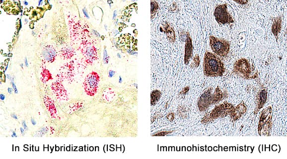
Dual RNAscope ISH-IHC Compatible
Sample: Immersion fixed paraffin-embedded sections of human placenta
Immunohistochemistry
Sample: Immersion fixed paraffin-embedded sections of human ovarian cancer tissue and normal human ovarian array
Knockout Validated
Western Blot
Sample: Recombinant Human TIMP-2 Western Blot Standard Protein (Catalog # WBC023), HeLa human cervical epithelial carcinoma cell line and human placenta tissue
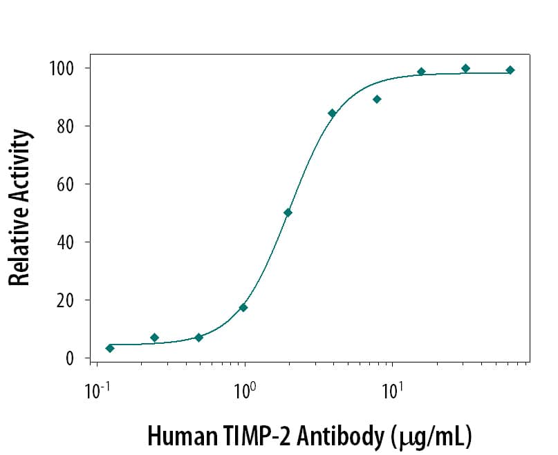
Neutralization
Reviewed Applications
Read 2 reviews rated 4 using AF971 in the following applications:
Formulation, Preparation, and Storage
Purification
Reconstitution
Formulation
*Small pack size (-SP) is supplied either lyophilized or as a 0.2 µm filtered solution in PBS.
Shipping
Stability & Storage
- 12 months from date of receipt, -20 to -70 °C as supplied.
- 1 month, 2 to 8 °C under sterile conditions after reconstitution.
- 6 months, -20 to -70 °C under sterile conditions after reconstitution.
Background: TIMP-2
Tissue inhibitors of metalloproteinases or TIMPs are a family of proteins that regulate the activation and proteolytic activity of the zinc enzymes known as matrix metalloproteinases (MMPs). There are four members of the family, TIMP-1, TIMP-2, TIMP-3, and TIMP-4. TIMP-2 is a non N-glycosylated protein with a molecular mass of 22 kDa produced by a wide range of cell types, which inhibits MMPs non-covalently by the formation of binary complexes. TIMP-2 also has erythroid-potentiating and cell growth promoting activities.
Long Name
Alternate Names
Gene Symbol
UniProt
Additional TIMP-2 Products
Product Documents for Human/Mouse TIMP-2 Antibody
Product Specific Notices for Human/Mouse TIMP-2 Antibody
For research use only


