Human Nestin Antibody
R&D Systems, part of Bio-Techne | Catalog # MAB1259

Conjugate
Catalog #
Key Product Details
Validated by
Biological Validation
Species Reactivity
Validated:
Human
Cited:
Human, Mouse, Rat, Drosophila, Xenograft
Applications
Validated:
CyTOF-reported, Immunocytochemistry, Intracellular Staining by Flow Cytometry
Cited:
Bioassay, Cell Culture, Flow Cytometry, Immunocytochemistry, Immunofluorescence, Immunohistochemistry, Immunohistochemistry-Frozen, Immunohistochemistry-Paraffin, Western Blot
Label
Unconjugated
Antibody Source
Monoclonal Mouse IgG1 Clone # 196908
Product Specifications
Immunogen
NS0 mouse myeloma cell line transfected with human Nestin
Specificity
Detects human Nestin.
Clonality
Monoclonal
Host
Mouse
Isotype
IgG1
Scientific Data Images for Human Nestin Antibody
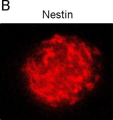
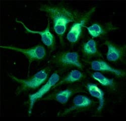
Nestin in Human Neural Progenitor Cells.
Nestin was detected in immersion fixed human fetal neural progenitor cells using 10 µg/mL Human Nestin Monoclonal Antibody (Catalog # MAB1259) for 3 hours at room temperature. Cells were stained (green) and counterstained with DAPI (blue). View our protocol for Fluorescent ICC Staining of Cells on Coverslips.Detection of Nestin in A172 cells by Flow Cytometry.
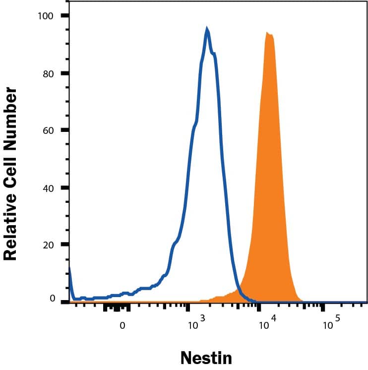
A172 cells were stained with Mouse Anti-Human Nestin Monoclonal Antibody (Catalog # MAB1259, filled histogram) or isotype control antibody (Catalog # MAB002, open histogram), followed by Phycoerythrin-conjugated Anti-Mouse IgG Secondary Antibody (Catalog # F0102B). To facilitate intracellular staining, cells were fixed and permeabilized with FlowX FoxP3 Fixation & Permeabilization Buffer Kit (Catalog # FC012). View our protocol for Staining Intracellular Molecules.Detection of Mouse Nestin by Western Blot
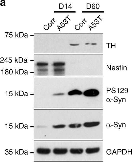
Cardiolipin is externalized to the mitochondrial surface in alpha-syn mutant hNs and transgenic mice in response to alpha-syn accumulation. a Western analysis of lysates from corrected and A53T cells at DIV 14 and DIV 60 labeled for total alpha-syn, PS129, TH (DA neuronal marker), nestin (NPC marker) or GAPDH show that levels of PS129-modified alpha-syn are elevated in A53T hNs at DIV 14 and remain elevated at DIV 60 relative to corrected cells. b, c Mitochondria were purified from A53T cells and genetically corrected cells at both DIV 14 and DIV 60. Lysates were then separated based on TX-100 solubility (soluble) or urea solubility (insoluble). Western analysis of A53T cells shows elevated soluble alpha-syn at the mitochondria relative to corrected cells at DIV 14 and DIV 60 (denoted by ** and * respectively). Quantification of soluble and insoluble alpha-syn levels in mitochondrial fractions normalized to Coomassie (c). Data represent mean ± s.e.m. *P < 0.05, **P < 0.01 by ANOVA followed by Tukey’s post hoc test, n = 4. d, e Micrographs of corrected and A53T-mutant NPCs expressing the negative charge probe RPRE-RFP and CL-GFP show RPRE-RFP translocation from a primarily plasma membrane localization in isogenic-corrected hNs to a punctate intracellular localization in A53T hNs in tight cellular localization with CL-GFP (d). Quantification of percent total cell number with colocalized RPRE-RFP and CL-GFP (e). Data represent mean ± s.e.m. **P < 0.0001 by t-test, n = 3 coverslips over 2 independent differentiations, DIV: 14; scale bar: 10 μm. f, g Fluorescence resonance energy transfer (FRET) from CL-GFP to RPRE-RFP in hiPSC-derived A53T and corrected hNs or hESC-derived WT, A53T and E46K hNs was assessed (f) and mean FRET intensity was quantified (g). Data represent mean ± s.e.m. **P < 0.01 by ANOVA followed by Tukey’s post hoc test, for hiPSCs, n = 6, for hESCs, n = 8 coverslips over two independent differentiations, DIV: 14. Scale bar: 10 µm. h Mitochondria were isolated from the SNpc of 6-month-old WT and A53T transgenic animals and labeled with Annexin V to measure the abundance of cardiolipin on the mitochondrial surface. Controls were rotenone-exposed WT samples. Animals were not randomized, nor was the analysis blinded. Data represent mean ± s.e.m. **P < 0.01 by ANOVA followed by Dunnett post hoc test, n = 4. Clipart was obtained at clker.com Image collected and cropped by CiteAb from the following publication (https://www.nature.com/articles/s41467-018-03241-9), licensed under a CC-BY license. Not internally tested by R&D Systems.Applications for Human Nestin Antibody
Application
Recommended Usage
CyTOF-reported
This clone has been commercially reported for use in CyTOF®. Ready to be labeled using established conjugation methods. No BSA or other carrier proteins that could interfere with conjugation.
Immunocytochemistry
8-25 µg/mL
Sample: Immersion fixed human neural progenitor cells
Sample: Immersion fixed human neural progenitor cells
Intracellular Staining by Flow Cytometry
0.25 µg/106 cells
Sample: A172 human glioblastoma cells fixed and permeabilized with FlowX FoxP3 Fixation & Permeabilization Buffer Kit (Catalog # FC012).
Sample: A172 human glioblastoma cells fixed and permeabilized with FlowX FoxP3 Fixation & Permeabilization Buffer Kit (Catalog # FC012).
Reviewed Applications
Read 7 reviews rated 4.3 using MAB1259 in the following applications:
Formulation, Preparation, and Storage
Purification
Protein A or G purified from hybridoma culture supernatant
Reconstitution
Reconstitute at 0.5 mg/mL in sterile PBS. For liquid material, refer to CoA for concentration.
Formulation
Lyophilized from a 0.2 μm filtered solution in PBS with Trehalose. *Small pack size (SP) is supplied either lyophilized or as a 0.2 µm filtered solution in PBS.
Shipping
Lyophilized product is shipped at ambient temperature. Liquid small pack size (-SP) is shipped with polar packs. Upon receipt, store immediately at the temperature recommended below.
Stability & Storage
Use a manual defrost freezer and avoid repeated freeze-thaw cycles.
- 12 months from date of receipt, -20 to -70 °C as supplied.
- 1 month, 2 to 8 °C under sterile conditions after reconstitution.
- 6 months, -20 to -70 °C under sterile conditions after reconstitution.
Background: Nestin
References
- Hockfield, S. and R.D. McKay (1985) J. Neurosci. 5:3310.
- Lendahl, U. et al. (1990) Cell 60:585.
- Frederiksen, K. and R.D. McKay (1988) J. Neurosci. 8:1144.
- Tohyama, T. et. al. (1992) Lab. Invest. 66:303.
- Uchida, N. et al. (2000) Proc. Natl. Acad.Sci. USA 97:14720.
- Frederiksen, K. et al. (1988) Neuron 1:439.
- Cattaneo, E. et al. (1990) Nature 347:762.
- Reynolds, B.A. and S. Weiss (1992) Science 255:1707.
- Rietze, R.L. et al. (2001) Nature 412:736.
- Carpenter, M.K. et al. (2001) Exp. Neurol 172:383.
- Zulewski, H. et al. (2001) Diabetes 50:521.
- Lumelsky, N. et al. (2001) Science 292:1389.
- Lechner, A. et al. (2002) Biochem.Biophys. Res. Commun. 293:670.
- Shih, C.C. et al. (2001) Blood 98:2412.
Alternate Names
NES
Gene Symbol
NES
Additional Nestin Products
Product Documents for Human Nestin Antibody
Product Specific Notices for Human Nestin Antibody
For research use only
Loading...
Loading...
Loading...
Loading...
Loading...
