Human PD-L1/B7-H1 Antibody
R&D Systems, part of Bio-Techne | Catalog # AF156

Key Product Details
Validated by
Species Reactivity
Validated:
Cited:
Applications
Validated:
Cited:
Label
Antibody Source
Product Specifications
Immunogen
Phe19-Thr239
Accession # Q9NZQ7
Specificity
Clonality
Host
Isotype
Scientific Data Images for Human PD-L1/B7-H1 Antibody
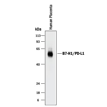
Detection of Human PD-L1/B7-H1 by Western Blot.
Western blot shows lysates of human placenta tissue. PVDF membrane was probed with 2 µg/mL of Goat Anti-Human PD-L1/B7-H1 Antigen Affinity-purified Polyclonal Antibody (Catalog # AF156) followed by HRP-conjugated Anti-Goat IgG Secondary Antibody (Catalog # HAF017). A specific band was detected for PD-L1/B7-H1 at approximately 50-55 kDa (as indicated). This experiment was conducted under reducing conditions and using Immunoblot Buffer Group 1.PD-L1/B7-H1 in Human Colon and Colon Cancer Tissue.
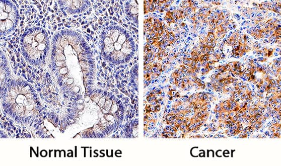
PD-L1/B7-H1 was detected in immersion fixed paraffin-embedded sections of normal human colon (left panel) and human colon cancer tissue (right panel) using Goat Anti-Human PD-L1/B7-H1 Antigen Affinity-purified Polyclonal Antibody (Catalog # AF156) at 5 µg/mL overnight at 4 °C. Tissue was stained using the Anti-Goat HRP-DAB Cell & Tissue Staining Kit (brown; Catalog # CTS008) and counterstained with hematoxylin (blue). Specific staining was localized to cell membranes and cytoplasm. View our protocol for Chromogenic IHC Staining of Paraffin-embedded Tissue Sections.Human PD-L1/B7-H1 ELISA Standard Curve.
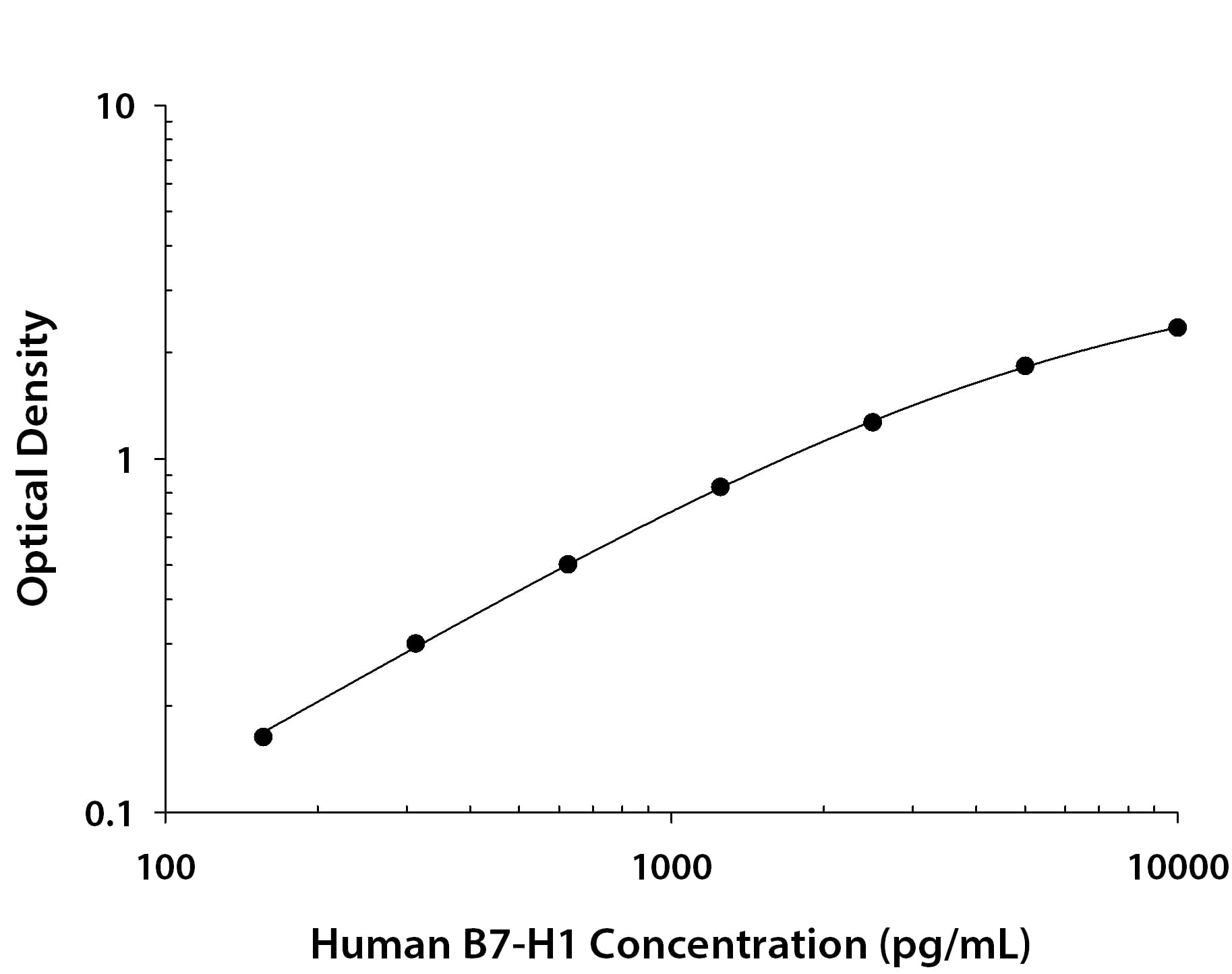
Recombinant Human PD-L1/B7-H1 protein was serially diluted 2-fold and captured by Mouse Anti-Human PD-L1/B7-H1 Monoclonal Antibody (Catalog # MAB1561R) coated on a Clear Polystyrene Microplate (Catalog # DY990). Goat Anti-Human PD-L1/B7-H1 Antigen Affinity-purified Polyclonal Antibody (Catalog # AF156) was biotinylated and incubated with the protein captured on the plate. Detection of the standard curve was achieved by incubating Streptavidin-HRP (Catalog # DY998) followed by Substrate Solution (Catalog # DY999) and stopping the enzymatic reaction with Stop Solution (Catalog # DY994).Applications for Human PD-L1/B7-H1 Antibody
ELISA
This antibody functions as an ELISA detection antibody when paired with Mouse Anti-Human PD-L1/B7-H1 Monoclonal Antibody (Catalog # MAB1561R).
This product is intended for assay development on various assay platforms requiring antibody pairs. We recommend the Human PD-L1/B7-H1 DuoSet ELISA Kit (Catalog # DY156) for convenient development of a sandwich ELISA or the Human/Cynomolgus Monkey PD-L1/B7-H1 Quantikine ELISA Kit (Catalog # DB7H10) for a complete optimized ELISA.
Immunohistochemistry
Sample: Immersion fixed paraffin-embedded sections of normal human colon and human colon cancer tissue
Simple Western
Sample: HDLM‑2 human Hodgkin's lymphoma cell line
Western Blot
Sample: Human placenta tissue
Reviewed Applications
Read 4 reviews rated 4.5 using AF156 in the following applications:
Formulation, Preparation, and Storage
Purification
Reconstitution
Formulation
Shipping
Stability & Storage
- 12 months from date of receipt, -20 to -70 °C as supplied.
- 1 month, 2 to 8 °C under sterile conditions after reconstitution.
- 6 months, -20 to -70 °C under sterile conditions after reconstitution.
Background: PD-L1/B7-H1
Human B7 homolog 1 (B7-H1), also called programmed cell death 1 ligand 1 (PDCD1L1) and programmed death ligand 1 (PDL1), is a member of the growing B7 family of immune proteins that provide signals for both stimulating and inhibiting T cell activation. Other family members include B7-1, B7-2, B7-H2, PDL2 and B7-H3. B7 proteins are members of the immunoglobulin (Ig) superfamily, their extracellular domains contain 2 Ig-like domains and all members have short cytoplasmic domains. Among the family members, they share about 20-25% amino acid identity. Human and mouse B7-H1 share approximately 70% amino acid sequence identity. B7-H1 has been identified as one of two ligands for programmed death-1 (PD-1), a member of the CD28 family of immunoreceptors. The B7-H1 gene encodes a 290 amino acid (aa) type I membrane precursor protein with a putative 18 aa signal peptide, a 221 aa extracellular domain, a 21 aa transmembrane region, and a 31 aa cytoplasmic domain. Human B7-H1 is constitutively expressed in several organs such as heart, skeletal muscle, placenta and lung, and in lower amounts in thymus, spleen, kidney and liver. B7-H1 expression is upregulated in a small fraction of activated T and B cells and a much larger fraction of activated monocytes. B7-H1 expression is also induced in dendritic cells and keratinocytes after IFN-gamma stimulation. Interaction of B7-H1 with PD-1 results in inhibition of TCR-mediated proliferation and cytokine production. The B7-H1:PD-1 pathway is involved in the negative regulation of some immune responses and may play an important role in the regulation of peripheral tolerance.
References
- Nishimura, H. and T. Honjo (2001) Trends in Immunology 22:265.
- Freeman, G.J. et al. (2000) J. Exp. Med. 192:1027.
- Latchman, Y. et al. (2001) Nat. Immunol. 2:261.
Long Name
Alternate Names
Entrez Gene IDs
Gene Symbol
UniProt
Additional PD-L1/B7-H1 Products
Product Documents for Human PD-L1/B7-H1 Antibody
Product Specific Notices for Human PD-L1/B7-H1 Antibody
For research use only
