Human STAT5a Antibody
R&D Systems, part of Bio-Techne | Catalog # MAB21741


Key Product Details
Species Reactivity
Applications
Label
Antibody Source
Product Specifications
Immunogen
SLDSRLSPPAGLFTSARGSLS
Accession # NP_003143
Specificity
Clonality
Host
Isotype
Scientific Data Images for Human STAT5a Antibody
Detection of STAT5a in Jurkat Human Cell Line by Flow Cytometry.
Jurkat human acute T cell leukemia cell line was stained with Mouse Anti-Human STAT5a Monoclonal Antibody (Catalog # MAB21741, filled histogram) or isotype control antibody (Catalog # MAB002, open histogram), followed by Allophycocyanin-conjugated Anti-Mouse IgG F(ab')2Secondary Antibody (Catalog # F0101B). To facilitate intracellular staining, cells were fixed with paraformaldehyde and permeabilized with methanol.STAT5a in K562 Human Cell Line.
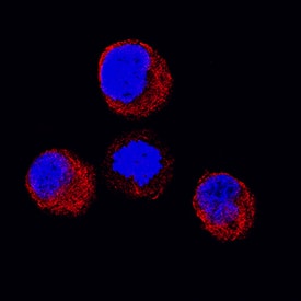
STAT5a was detected in immersion fixed K562 human chronic myelogenous leukemia cell line using Mouse Anti-Human STAT5a Monoclonal Antibody (Catalog # MAB21741) at 25 µg/mL for 3 hours at room temperature. Cells were stained using the NorthernLights™ 557-conjugated Anti-Mouse IgG Secondary Antibody (red; Catalog # NL007) and counterstained with DAPI (blue). Specific staining was localized to cytoplasm. View our protocol for Fluorescent ICC Staining of Cells on Coverslips.Applications for Human STAT5a Antibody
CyTOF-ready
Immunocytochemistry
Sample: Immersion fixed K562 human chronic myelogenous leukemia cell line and human peripheral blood mononuclear cells
Intracellular Staining by Flow Cytometry
Sample: Jurkat human acute T cell leukemia cell line fixed with paraformaldehyde and permeabilized with methanol
Reviewed Applications
Read 3 reviews rated 4 using MAB21741 in the following applications:
Formulation, Preparation, and Storage
Purification
Reconstitution
Formulation
Shipping
Stability & Storage
- 12 months from date of receipt, -20 to -70 °C as supplied.
- 1 month, 2 to 8 °C under sterile conditions after reconstitution.
- 6 months, -20 to -70 °C under sterile conditions after reconstitution.
Background: STAT5a
STAT5a (Signal Transducer and Activator of Transcription-5a) is one of two closely related genes that belong to the STAT family of transcription factors. It is a 91 kDa cytosolic protein that contains an N-terminal domain (with an NES/Nuclear Export Signal) a coiled-coiled region (with an NLS/Nuclear Localization Signal) a DNA-binding site that recognizes a GAS (Gamma-interferon Activated Site) motif, an SH2 domain that allows for dimerization, and a C-terminal transactivation domain. STAT5a likely exists in a quiescent state as a cytoplasmic anti-parallel homodimer. Following activation of both tyrosine and non-tyrosine kinase membrane-bound receptors, STAT5a is phosphorylated, increasing its MW by some 5-6 kDa. Phosphorylated STAT5a will enter the nucleus as either a homodimer (or heterodimer with STAT5b), or as a complex with the intracellular domain of a tyrosine kinase receptor such as ErbB4. Once in the nucleus, STAT5a will form a homotetramer and bind either GAS or GAS-related sequences in gene regulatory regions. Notably, it is suggested that STAT5a may "cycle" through the nucleus without phosphorylation, a process that would seem to involve Importins alpha3 and beta1. STAT5a is related to STAT5b through gene duplication. They show 92% amino acid (aa) sequence identity, with the major differences existing over aa 45-56 and 773-794. The genes are not entirely redundant; STAT5a activates NDGR1 and SH2, while STAT5b regulates the Treg T cell genes, FoxP3 and CD25/IL-2R alpha.
Long Name
Alternate Names
Gene Symbol
UniProt
Additional STAT5a Products
Product Documents for Human STAT5a Antibody
Product Specific Notices for Human STAT5a Antibody
For research use only
