Human VEGFR2/KDR/Flk-1 Antibody
R&D Systems, part of Bio-Techne | Catalog # MAB3572


Key Product Details
Species Reactivity
Validated:
Cited:
Applications
Validated:
Cited:
Label
Antibody Source
Product Specifications
Immunogen
Ala20-Glu764
Accession # P35968
Specificity
Clonality
Host
Isotype
Endotoxin Level
Scientific Data Images for Human VEGFR2/KDR/Flk-1 Antibody
Detection of VEGFR2/KDR/Flk‑1 in HUVEC Human Cells by Flow Cytometry.
HUVEC human umbilical vein endothelial cells were stained with Mouse Anti-Human VEGFR2/KDR/Flk-1 Monoclonal Antibody (Catalog # MAB3572, filled histogram) or isotype control antibody (Catalog # MAB002, open histogram), followed by Phycoerythrin-conjugated Anti-Mouse IgG Secondary Antibody (Catalog # F0102B).VEGFR2/KDR Inhibition of VEGF-dependent Cell Proliferation and Neutralization by Human VEGFR2/KDR Antibody.
Recombinant Human VEGFR2/KDR Fc Chimera (Catalog # 357-KD) inhibits Recombinant Human VEGF165(Catalog # 293-VE) induced proliferation in HUVEC human umbilical vein endothelial cells in a dose-dependent manner (orange line). Inhibition of Recombinant Human VEGF165(10 ng/mL) activity elicited by Recombinant Human VEGFR2/KDR Fc Chimera (50 ng/mL) is neutralized (green line) by increasing concentrations of Mouse Anti-Human VEGFR2/KDR Monoclonal Antibody (Catalog # MAB3572). The ND50 is typically 10-50 ng/mL.Detection of VEGFR2/KDR/Flk-1 by Flow Cytometry
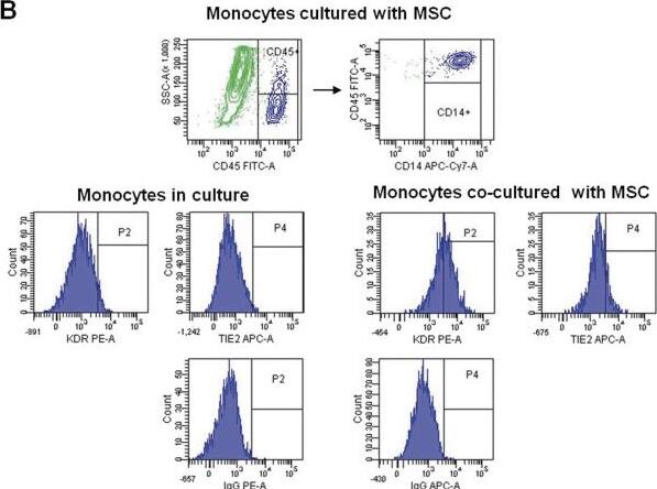
Induction of endothelial markers in circulating CD14+ cells. CD14+ monocytes were isolated from adult PB, cultured in EGM-2 MV with or without MSCs, and then analyzed with flow cytometry. Dot plots from freshly isolated CD14+ cells (A) and after 4 days of co-culture with adipose-derived MSCs (green) (B) are shown. Flow-cytometry histograms in panels A and B show the expression of KDR and Tie2 in gated CD14+ cells. Isotype-matched controls are given. Image collected and cropped by CiteAb from the following open publication (https://pubmed.ncbi.nlm.nih.gov/24731246), licensed under a CC-BY license. Not internally tested by R&D Systems.Applications for Human VEGFR2/KDR/Flk-1 Antibody
CyTOF-ready
Flow Cytometry
Sample: HUVEC human umbilical vein endothelial cells
Neutralization
Reviewed Applications
Read 2 reviews rated 5 using MAB3572 in the following applications:
Formulation, Preparation, and Storage
Purification
Reconstitution
Formulation
Shipping
Stability & Storage
- 12 months from date of receipt, -20 to -70 °C as supplied.
- 1 month, 2 to 8 °C under sterile conditions after reconstitution.
- 6 months, -20 to -70 °C under sterile conditions after reconstitution.
Background: VEGFR2/KDR/Flk-1
VEGFR2 (KDR/Flk-1), VEGFR1 (Flt-1) and VEGFR3 (Flt-4) belong to the class III subfamily of receptor tyrosine kinases (RTKs). All three receptors contain seven immunoglobulin-like repeats in their extracellular domains and kinase insert domains in their intracellular regions. The expression of VEGFR1, 2, and 3 is almost exclusively restricted to the endothelial cells. These receptors are likely to play essential roles in vasculogenesis and angiogenesis. Mature VEGFR2 is composed of a 745 aa extracellular domain, a 25 aa transmembrane domain and a 567 aa cytoplasmic domain. In contrast to VEGFR1 which binds both PlGF and VEGF with high affinity, VEGFR2 binds VEGF but not PlGF with high affinity. The recombinant soluble VEGFR2/Fc chimera binds VEGF with high affinity and is a potent VEGF antagonist.
References
- Ferra, N. and R. Davis-Smyth (1997) Endocrine Reviews 18:4.
Long Name
Alternate Names
Gene Symbol
UniProt
Additional VEGFR2/KDR/Flk-1 Products
Product Documents for Human VEGFR2/KDR/Flk-1 Antibody
Product Specific Notices for Human VEGFR2/KDR/Flk-1 Antibody
For research use only

