Mouse LYVE-1 Antibody Best Seller
R&D Systems, part of Bio-Techne | Catalog # AF2125


Key Product Details
Species Reactivity
Validated:
Cited:
Applications
Validated:
Cited:
Label
Antibody Source
Product Specifications
Immunogen
Ala24-Thr234
Accession # Q8BHC0
Specificity
Clonality
Host
Isotype
Scientific Data Images for Mouse LYVE-1 Antibody
LYVE-1 in Mouse Liver.
LYVE-1 was detected in perfusion fixed frozen sections of mouse liver using 15 µg/mL Goat Anti-Mouse LYVE-1 Antigen Affinity-purified Polyclonal Antibody (Catalog # AF2125) overnight at 4 °C. Tissue was stained with the Anti-Goat HRP-DAB Cell & Tissue Staining Kit (brown; CTS008) and counterstained with hematoxylin (blue). Specific labeling was localized to the cytoplasm of endothelial cells in sinusoids. View our protocol for Chromogenic IHC Staining of Frozen Tissue Sections.Detection of Mouse LYVE‑1 by Western Blot.
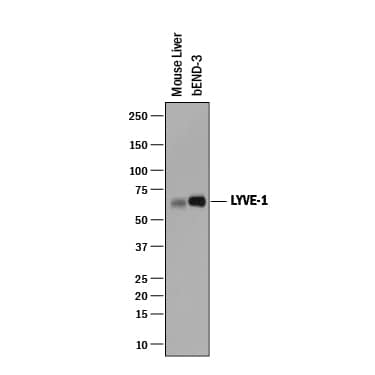
Western blot shows lysates of mouse liver tissue and bEnd.3 mouse brain endothelial cell line. PVDF membrane was probed with 0.25 µg/mL of Goat Anti-Mouse LYVE-1 Antigen Affinity-purified Polyclonal Antibody (Catalog # AF2125) followed by HRP-conjugated Anti-Goat IgG Secondary Antibody (HAF019). A specific band was detected for LYVE-1 at approximately 60-65 kDa (as indicated). This experiment was conducted under reducing conditions and using Immunoblot Buffer Group 1.Detection of Mouse LYVE-1 by Immunocytochemistry/Immunofluorescence
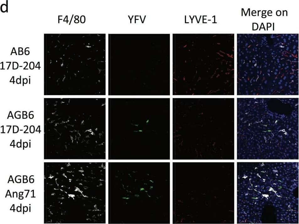
17D-204 causes viscerotropic abnormalities in the absence of IFN-gamma. H&E sections (a–c) antibody-stained frozen sections (b, d) of spleen (a, b) and liver (c, d) were presented. a Spleen sections from 17D-204-infected animals were indistinguishable at 4 dpi, whereas Angola71-infected animals display loss of white pulp and red pulp architecture and increase in infiltrating macrophages and neutrophils (inset) at 4 dpi. At 11 dpi, 17D-204 infected AGB6 has increased immune infiltration and extramedullary hematopoiesis (inset) but not AB6 mice. b YFV antigen can be detected in the spleen at 4 dpi in both AB6 and AGB6 mice in the red pulp and outer marginal zone area. c 17D-204-infected AB6 mice do not display major histological changes but infected AGB6 mice and Angola71-infected animals had microsteatosis (inset) at 4 dpi. In AGB6 mice, microsteatosis only occurs transiently at 4 dpi and resolved by 11 dpi. Antibody-stained frozen liver sections (d) revealed that YFV antigen could only be detected in infected AGB6 mice at 4 dpi but not AB6 mice, parallel to titer data in Fig. 1g. Original magnification = 40x, n = 4 Image collected and cropped by CiteAb from the following publication (https://www.nature.com/articles/s41541-017-0039-z), licensed under a CC-BY license. Not internally tested by R&D Systems.Applications for Mouse LYVE-1 Antibody
Dual RNAscope ISH-IHC Compatible
Sample: Immersion fixed paraffin-embedded sections of mouse liver
Immunohistochemistry
Sample: Perfusion fixed frozen sections of mouse liver
Western Blot
Sample: Mouse liver tissue and bEnd.3 mouse brain endothelial cell line
Reviewed Applications
Read 4 reviews rated 4.8 using AF2125 in the following applications:
Formulation, Preparation, and Storage
Purification
Reconstitution
Formulation
*Small pack size (-SP) is supplied either lyophilized or as a 0.2 µm filtered solution in PBS.
Shipping
Stability & Storage
- 12 months from date of receipt, -20 to -70 °C as supplied.
- 1 month, 2 to 8 °C under sterile conditions after reconstitution.
- 6 months, -20 to -70 °C under sterile conditions after reconstitution.
Background: LYVE-1
Lymphatic vessel endothelial hyaluronan (HA) receptor-1 (LYVE-1) is a recently identified receptor of HA, a linear high molecular weight polymer composed of alternating units of D-glucuronic acid and N-acetyl-D-glucosamine. HA is found in the extracellular matrix of most animal tissues and in body fluids. It modulates cell behavior and functions during tissue remodeling, development, homeostasis, and disease. The turnover of HA (several grams/day in humans) occurs primarily in the lymphatics and liver, the two major clearance systems that catabolize approximately 85% and 15% of HA, respectively. LYVE-1 shares 41% homology with the other known HA receptor, CD44. The homology between the two proteins increases to 61% within the HA binding domain. The HA binding domain, known as the link module, is a common structural motif found in other HA binding proteins such as link protein, aggrecan and versican. Human and mouse LYVE-1 share 69% amino acid sequence identity.
LYVE-1 is primarily expressed on both the luminal and abluminal surfaces of lymphatic vessels. In addition, LYVE-1 is also present in normal hepatic blood sinusoidal endothelial cells. LYVE-1 mediates the endocytosis of HA and may transport HA from tissue to lymph by transcytosis, delivering HA to lymphatic capillaries for removal and degradation in the regional lymph nodes. Because of its restricted expression patterns, LYVE-1, along with other lymphatic proteins such as VEGF R3, podoplanin and the homeobox protein propero-related (Prox-1), constitute a set of markers useful for distinguishing between lymphatic and blood microvasculature.
Long Name
Alternate Names
Gene Symbol
UniProt
Additional LYVE-1 Products
Product Documents for Mouse LYVE-1 Antibody
Product Specific Notices for Mouse LYVE-1 Antibody
For research use only


