Mouse/Rat CD200 Antibody
R&D Systems, part of Bio-Techne | Catalog # AF3355


Key Product Details
Validated by
Species Reactivity
Validated:
Cited:
Applications
Validated:
Cited:
Label
Antibody Source
Product Specifications
Immunogen
Gln31-Gly232
Accession # O54901
Specificity
Clonality
Host
Isotype
Scientific Data Images for Mouse/Rat CD200 Antibody
Detection of Mouse and Rat CD200 by Western Blot.
Western blot shows lysates of mouse brain tissue and rat brain tissue. PVDF membrane was probed with 0.25 µg/mL of Goat Anti-Mouse/Rat CD200 Antigen Affinity-purified Polyclonal Antibody (Catalog # AF3355) followed by HRP-conjugated Anti-Goat IgG Secondary Antibody (Catalog # HAF019). A specific band was detected for CD200 at approximately 38-45 kDa (as indicated). This experiment was conducted under reducing conditions and using Immunoblot Buffer Group 1.CD200 in Mouse Brain.
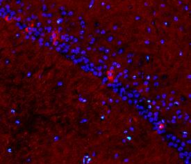
CD200 was detected in perfusion fixed frozen sections of normal mouse brain using Goat Anti-Mouse/Rat CD200 Antigen Affinity-purified Polyclonal Antibody (Catalog # AF3355) at 15 µg/mL overnight at 4 °C. Tissue was stained using the NorthernLights™ 557-conjugated Anti-Goat IgG Secondary Antibody (red; Catalog # NL001) and counterstained with DAPI (blue). Specific staining was localized to plasma membranes of hippocampal neurons. View our protocol for Fluorescent IHC Staining of Frozen Tissue Sections.Detection of Mouse and Rat CD200 by Simple WesternTM.
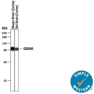
Simple Western lane view shows lysates of mouse brain (cortex) tissue and rat brain (cortex) tissue, loaded at 0.2 mg/mL. A specific band was detected for CD200 at approximately 73-76 kDa (as indicated) using 2.5 µg/mL of Goat Anti-Mouse/Rat CD200 Antigen Affinity-purified Polyclonal Antibody (Catalog # AF3355) followed by 1:50 dilution of HRP-conjugated Anti-Goat IgG Secondary Antibody (Catalog # HAF109). This experiment was conducted under reducing conditions and using the 12-230 kDa separation system.Applications for Mouse/Rat CD200 Antibody
Immunohistochemistry
Sample: Perfusion fixed frozen sections of mouse brain (cortex)
Simple Western
Sample: Mouse brain (cortex) tissue and rat brain (cortex) tissue
Western Blot
Sample: Mouse brain tissue and rat brain tissue
Formulation, Preparation, and Storage
Purification
Reconstitution
Formulation
Shipping
Stability & Storage
- 12 months from date of receipt, -20 to -70 °C as supplied.
- 1 month, 2 to 8 °C under sterile conditions after reconstitution.
- 6 months, -20 to -70 °C under sterile conditions after reconstitution.
Background: CD200
CD200, also known as OX-2, is a 45 kDa type I transmembrane immunoregulatory protein that belongs to the immunoglobulin superfamily (1, 2). The mouse CD200 cDNA encodes a 278 amino acid (aa) precursor that includes a 30 aa signal sequence, a 202 aa extracellular domain (ECD), a 27 aa transmembrane segment, and a 19 aa cytoplasmic domain. The ECD is composed of one Ig-likeV-type and one Ig-like C2-type domain (3). Splice variants of CD200 have been described in human but not in mouse. Within the ECD, mouse CD200 shares 76% and 94% aa sequence identity with human and rat CD200, respectively. CD200 is widely but not ubiquitously expressed (4). Its receptor (CD200R) is restricted primarily to mast cells, basophils, macrophages, and dendritic cells, which suggests myeloid cell regulation as the major function of CD200 (5‑7). CD200 knockout mice are characterized by increased macrophage number and activation, and are predisposed to autoimmune disorders (8). CD200 and CD200 R associate via their respective N-terminal Ig-like domains (9). In myeloid cells, CD200 R initiates inhibitory signals following receptor-ligand contact (6, 7, 10). In T cells, CD200 functions as a costimulatory molecule that is independent of the CD28 pathway (11). Several additional CD200 R-like molecules have been identified in human and mouse, but their capacity to interact with CD200 is controversial (12, 13). Several viruses encode CD200 homologs which are expressed on infected cells during the lytic phase (14, 15). Like CD200 itself, viral CD200 homologs also suppress myeloid cell activity, enabling increased viral propagation (5, 14‑16).
References
- Gorczynski, R.M. (2005) Curr. Opin. Invest. Drugs 6:483.
- Barclay, A.N. et al. (2002) Trends Immunol. 23:285.
- Chen, Z. et al. (1997) Biochim. Biophys. Acta 1362:6.
- Wright, G.J. et al. (2001) Immunology 102:173.
- Shiratori, I. et al. (2005) J. Immunol. 175:4441.
- Cherwinski, H.M. et al. (2005) J. Immunol. 174:1348.
- Fallarino, F. et al. (2004) J. Immunol. 173:3748.
- Hoek, R.M. et al. (2000) Science 290:1768.
- Hatherley, D. and A.N. Barclay (2004) Eur. J. Immunol. 34:1688.
- Jenmalm, M.C. et al. (2006) J. Immunol. 176:191.
- Borriello, F. et al. (1997) J. Immunol. 158:4548.
- Gorczynski, R. et al. (2004) J. Immunol. 172:7744.
- Hatherley, D. et al. (2005) J. Immunol. 175:2469.
- Foster-Cuevas, M. et al. (2004) J. Virol. 78:7667.
- Cameron, C.M. et al. (2005) J. Virol. 79:6052.
- Langlais, C.L. et al. (2006) J. Virol. 80:3098.
Alternate Names
Gene Symbol
UniProt
Additional CD200 Products
Product Documents for Mouse/Rat CD200 Antibody
Product Specific Notices for Mouse/Rat CD200 Antibody
For research use only
