Recombinant Mouse PD-L1/B7-H1 Fc Chimera Protein, CF
R&D Systems, part of Bio-Techne | Catalog # 1019-B7

Key Product Details
Source
NS0
Accession #
Structure / Form
Disulfide-linked homodimer
Conjugate
Unconjugated
Applications
Bioactivity
Product Specifications
Source
Mouse myeloma cell line, NS0-derived mouse PD-L1/B7-H1 protein
| Mouse PD-L1 (Phe19-Thr238) Accession # Q9EP73 |
IEGRMD | Human IgG1 (Pro100-Lys330) |
| N-terminus | C-terminus |
Purity
>90%, by SDS-PAGE visualized with Silver Staining and quantitative densitometry by Coomassie® Blue Staining.
Endotoxin Level
<0.10 EU per 1 μg of the protein by the LAL method.
N-terminal Sequence Analysis
Phe19
Predicted Molecular Mass
51.3 kDa (monomer)
SDS-PAGE
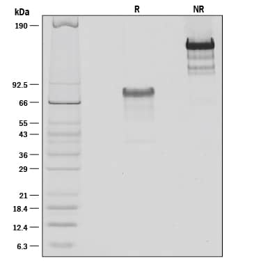
75-85 kDa, reducing conditions
Activity
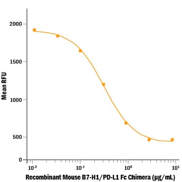
Measured by its ability to inhibit anti-CD3-induced proliferation of stimulated mouse T cells.
The ED50 for this effect is 0.15-0.75 µg/mL.
The ED50 for this effect is 0.15-0.75 µg/mL.
Reviewed Applications
Read 13 reviews rated 5 using 1019-B7 in the following applications:
Scientific Data Images for Recombinant Mouse PD-L1/B7-H1 Fc Chimera Protein, CF
Recombinant Mouse PD-L1/B7-H1 Fc Chimera Protein Bioactivity
Recombinant Mouse PD-L1/B7-H1 Fc Chimera (Catalog # 1019-B7) inhibits anti-CD3-induced cell proliferation of stimulated mouse T cells. The ED50 for this effect is 0.15-0.75 μg/mL.Recombinant Mouse PD-L1/B7-H1 Fc Chimera Protein SDS-PAGE
1 μg/lane of Recombinant Mouse PD-L1/B7-H1 Fc Chimera was resolved with SDS-PAGE under reducing (R) and non-reducing (NR) conditions and visualized by silver staining, showing bands at 79 kDa and 150 kDa, respectively.Binding of Mouse PD-1 to PD-L1/B7-H1 by surface plasmon resonance (SPR).
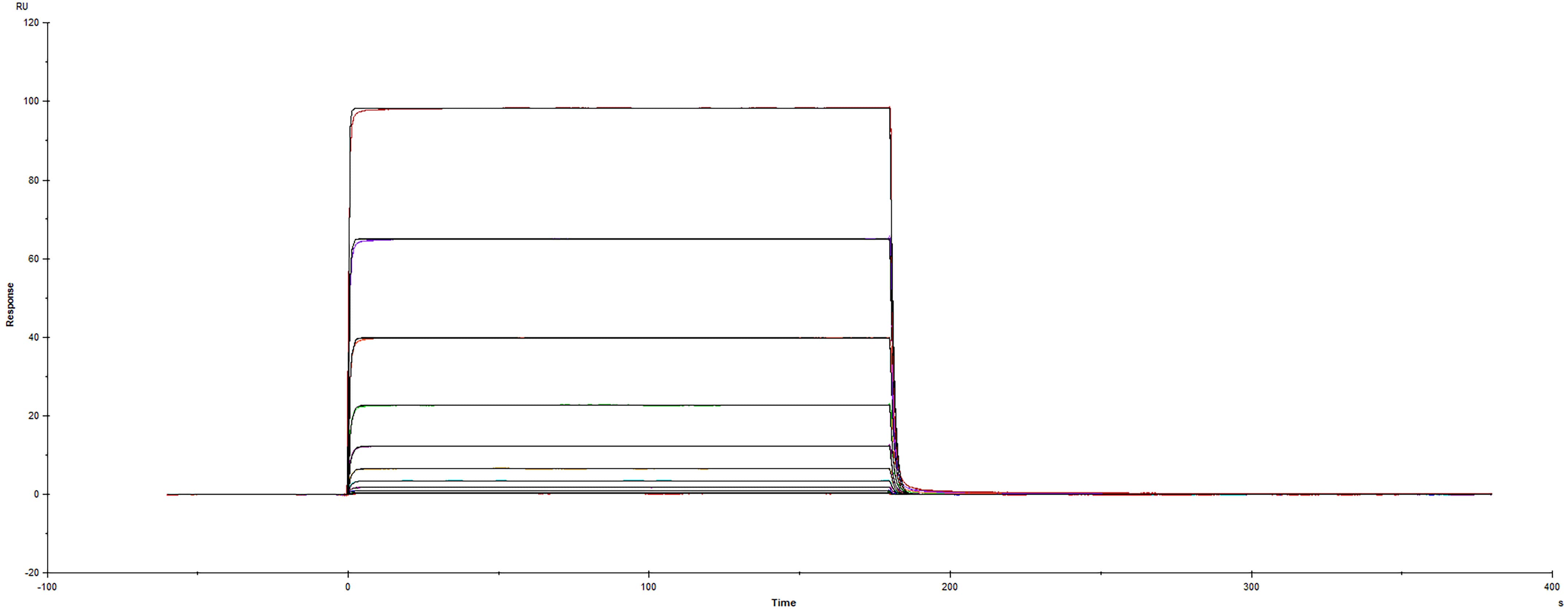
Recombinant Mouse PD-L1/B7-H1 Fc protein (Catalog # 1019-B7) was immobilized on a Biacore Sensor Chip CM5, and binding to Recombinant Mouse PD-1 His protein (9047-PD) was measured at a concentration range between 1.53 nM and 1.56 uM. The double-referenced sensorgram was fit to a 1:1 binding model to determine the binding kinetics and affinity, with an affinity constant of KD=0.539 uM.Formulation, Preparation and Storage
1019-B7
| Formulation | Lyophilized from a 0.2 μm filtered solution in PBS. |
| Reconstitution |
Reconstitute at 100 μg/mL in sterile PBS.
|
| Shipping | The product is shipped at ambient temperature. Upon receipt, store it immediately at the temperature recommended below. |
| Stability & Storage | Use a manual defrost freezer and avoid repeated freeze-thaw cycles.
|
Background: PD-L1/B7-H1
References
- Ceeraz, S. et al. (2013) Trends Immunol. 34:556.
- Tamura, H. et al. (2001) Blood 97:1809.
- Chen, L. et al. (2007) J. Immunol. 178:6634.
- Kuang, D.-M. et al. (2014) J. Clin. Invest. 124:4657.
- Tsushima, F. et al. (2007) Blood 110:180.
- Mazanet, M.M. and C.C.W. Hughes (2002) J. Immunol. 169:3581.
- Cao, Y. et al. (2010) Cancer Res. 71:1235.
- Scandiuzzi, L. et al. (2014) Cell Rep. 6:625.
- Dong, H. et al. (2002) Nat. Med. 8:793.
- Azuma, T. et al. (2008) Blood 111:3635.
- Butte, M.J. et al. (2008) Mol. Immunol. 45:3567.
- Park, J.-J. et al. (2010) Blood 116:1291.
- Ritprajak, P. et al. (2010) J. Immunol. 184:4918.
- Herold, M. et al. (2015) J. Immunol. 195:3584.
Long Name
Programmed Death Ligand 1
Alternate Names
B7-H1, B7H1, CD274, PDCD1L1, PDCD1LG1, PDL1
Entrez Gene IDs
Gene Symbol
CD274
UniProt
Additional PD-L1/B7-H1 Products
Product Documents for Recombinant Mouse PD-L1/B7-H1 Fc Chimera Protein, CF
Product Specific Notices for Recombinant Mouse PD-L1/B7-H1 Fc Chimera Protein, CF
For research use only
Loading...
Loading...
Loading...
Loading...


