Human c-Myc Antibody Best Seller
R&D Systems, part of Bio-Techne | Catalog # MAB3696

Key Product Details
Validated by
Species Reactivity
Validated:
Cited:
Applications
Validated:
Cited:
Label
Antibody Source
Product Specifications
Immunogen
Ala408-Ala439
Accession # P01106
Specificity
Clonality
Host
Isotype
Scientific Data Images for Human c-Myc Antibody
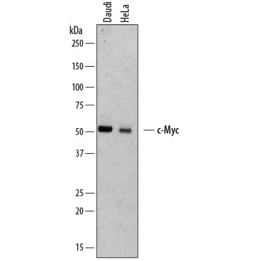
Detection of Human c‑Myc by Western Blot.
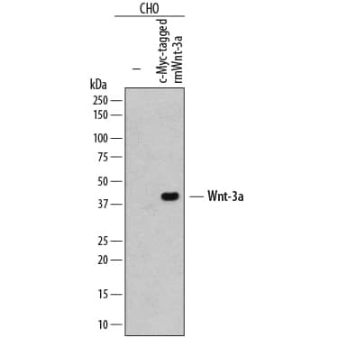
Western blot shows lysates of Daudi human Burkitt's lymphoma cell line and HeLa human cervical epithelial carcinoma cell line. PVDF membrane was probed with 2 µg/mL of Mouse Anti-Human c-Myc Monoclonal Antibody (Catalog # MAB3696) followed by HRP-conjugated Anti-Mouse IgG Secondary Antibody (Catalog # HAF007). A specific band was detected for c-Myc at approximately 52 kDa (as indicated). This experiment was conducted under reducing conditions and using Immunoblot Buffer Group 1.Detection of c‑Myc-tagged Protein by Western Blot.
Western blot shows lysates of CHO Chinese hamster ovary cell line either mock transfected (-) or transfected with c-Myc-tagged recombinant mouse Wnt-3a. PVDF membrane was probed with 2 µg/mL of Mouse Anti-Human c-Myc Monoclonal Antibody (Catalog # MAB3696) followed by HRP-conjugated Anti-Mouse IgG Secondary Antibody (Catalog # HAF007). A specific band was detected for c-Myc-tagged recombinant mouse Wnt-3a at approximately 41 kDa (as indicated). This experiment was conducted under reducing conditions and using Immunoblot Buffer Group 1.Detection of c‑Myc in Jurkat Human Cell Line by Flow Cytometry.
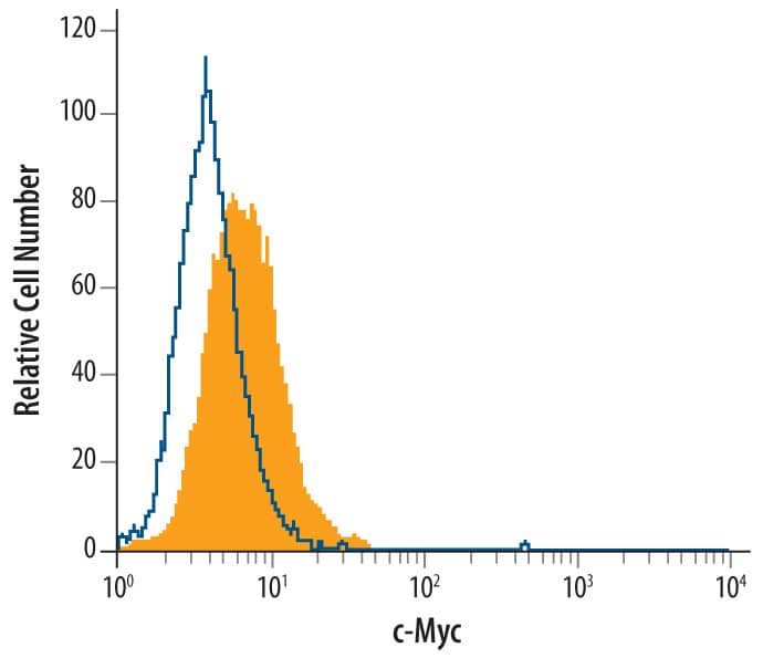
Jurkat human acute T cell leukemia cell line was stained with Mouse Anti-Human c-Myc Mono-clonal Antibody (Catalog # MAB3696, filled histogram) or isotype control antibody (Catalog # MAB002, open histogram), followed by Phycoerythrin-conjugated Anti-Mouse IgG Secondary Antibody (Catalog # F0102B). To facilitate intracellular staining, cells were fixed with paraformaldehyde and permeabilized with methanol.Applications for Human c-Myc Antibody
CyTOF-ready
Flow Cytometry
Sample: Jurkat human acute T cell leukemia cell line fixed with paraformaldehyde and permeabilized with methanol.
Immunocytochemistry
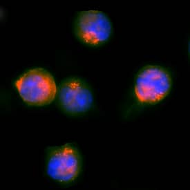
Sample: Immersion fixed HEK293 human embryonic kidney cell line
Immunohistochemistry
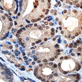
Sample: Immersion fixed paraffin-embedded sections of human prostate
Immunoprecipitation
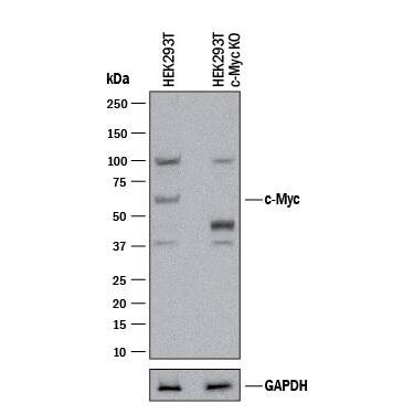
Knockout Validated
Simple Western
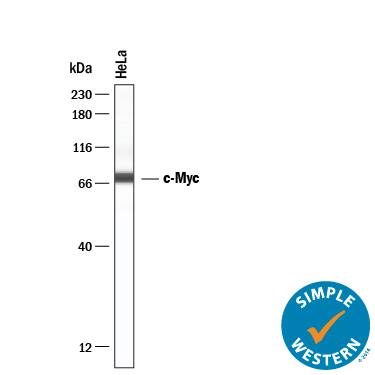
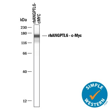
Sample: HeLa human cervical epithelial carcinoma cell line and c-Myc-tagged recombinant human ANGPTL6
Western Blot
Sample: Daudi human Burkitt's lymphoma cell line, HeLa human cervical epithelial carcinoma cell line, and CHO Chinese hamster ovary cell line either transfected with c-Myc-tagged recombinant mouse Wnt-3a
Reviewed Applications
Read 3 reviews rated 4.3 using MAB3696 in the following applications:
Formulation, Preparation, and Storage
Purification
Reconstitution
Formulation
Shipping
Stability & Storage
- 12 months from date of receipt, -20 to -70 °C as supplied.
- 1 month, 2 to 8 °C under sterile conditions after reconstitution.
- 6 months, -20 to -70 °C under sterile conditions after reconstitution.
Background: c-Myc
Human c-Myc is a helix-loop-helix transcription factor which efficiently binds DNA after heterodimerization with the bHLH protein Max. It is often overexpressed and mutated in hematopoietic tumors. Mutations frequently result in truncation around amino acid (aa) 252, before the C-terminal DNA binding, HLH and leucine zipper domains. The 439 aa human c-Myc has one O-glycosylation site and has three Ser/Thr phosphorylation sites near the N-terminus. Human c-Myc shows 92% aa identity with mouse or rat c-Myc.
Long Name
Alternate Names
Gene Symbol
UniProt
Additional c-Myc Products
Product Documents for Human c-Myc Antibody
Product Specific Notices for Human c-Myc Antibody
For research use only
