Human Integrin alpha3/CD49c Antibody
R&D Systems, part of Bio-Techne | Catalog # MAB1345


Conjugate
Catalog #
Key Product Details
Species Reactivity
Validated:
Human
Cited:
Human
Applications
Validated:
CyTOF-ready, Flow Cytometry, Immunocytochemistry
Cited:
Flow Cytometry, Functional Assay, Immunohistochemistry, Neutralization
Label
Unconjugated
Antibody Source
Monoclonal Mouse IgG1 Clone # IA3
Product Specifications
Immunogen
Human milk epithelial cell line
Specificity
Detects human Integrin alpha3/CD49c.
Clonality
Monoclonal
Host
Mouse
Isotype
IgG1
Scientific Data Images for Human Integrin alpha3/CD49c Antibody
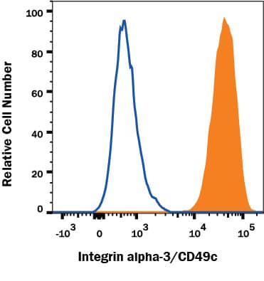
Detection of Integrin alpha 3/CD49c in HT1080 Human Cell Line by Flow Cytometry.
HT1080 human fibrosarcoma cell line was stained with Mouse Anti-Human Integrin alpha 3/CD49c Monoclonal Antibody (Catalog # MAB1345, filled histogram) or isotype control antibody (Catalog # MAB002, open histogram) followed by anti-Mouse IgG PE-conjugated secondary antibody (Catalog # F0102B). View our protocol for Staining Membrane-associated Proteins.Integrin alpha3/CD49c in HT1080 Human Cell Line.
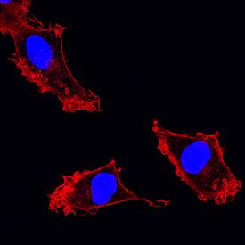
Integrin a3/CD49c was detected in immersion fixed HT1080 human fibrosarcoma cell line using Mouse Anti-Human Integrin a3/CD49c Monoclonal Antibody (Catalog # MAB1345) at 25 µg/mL for 3 hours at room temperature. Cells were stained using the NorthernLights™ 557-conjugated Anti-Mouse IgG Secondary Antibody (red; Catalog # NL007) and counterstained with DAPI (blue). Specific staining was localized to plasma membranes and cytoplasm. View our protocol for Fluorescent ICC Staining of Cells on Coverslips.Applications for Human Integrin alpha3/CD49c Antibody
Application
Recommended Usage
CyTOF-ready
Ready to be labeled using established conjugation methods. No BSA or other carrier proteins that could interfere with conjugation.
Flow Cytometry
0.25 µg/106 cells
Sample: HT1080 human fibrosarcoma cell line
Sample: HT1080 human fibrosarcoma cell line
Immunocytochemistry
8-25 µg/mL
Sample: Immersion fixed HT1080 human fibrosarcoma cell line
Sample: Immersion fixed HT1080 human fibrosarcoma cell line
Reviewed Applications
Read 2 reviews rated 4 using MAB1345 in the following applications:
Formulation, Preparation, and Storage
Purification
Protein A or G purified from hybridoma culture supernatant
Reconstitution
Reconstitute at 0.5 mg/mL in sterile PBS. For liquid material, refer to CoA for concentration.
Formulation
Lyophilized from a 0.2 μm filtered solution in PBS with Trehalose. *Small pack size (SP) is supplied either lyophilized or as a 0.2 µm filtered solution in PBS.
Shipping
Lyophilized product is shipped at ambient temperature. Liquid small pack size (-SP) is shipped with polar packs. Upon receipt, store immediately at the temperature recommended below.
Stability & Storage
Use a manual defrost freezer and avoid repeated freeze-thaw cycles.
- 12 months from date of receipt, -20 to -70 °C as supplied.
- 1 month, 2 to 8 °C under sterile conditions after reconstitution.
- 6 months, -20 to -70 °C under sterile conditions after reconstitution.
Background: Integrin alpha 3/CD49c
References
- Tsuji, T. et al. (2004) J. Membr. Biol. 200:115.
- Gu, J. and N. Taniguchi (2004) Glycoconj. J. 21:9.
- Kreidberg, J.A. (2000) Curr. Opin. Cell Biol. 12:548.
- Takada, Y. et al. (1991) J. Cell. Biol. 115:257.
- de Melker, A.A. et al. (1997) Lab. Invest. 76:547.
- Krokhin, O.V. et al. (2003) Biochemistry 42:12950.
- Springer, T.A. (2002) Curr. Opin. Struct. Biol. 12:802.
- Nishiuchi, R. et al. (2005) Proc. Natl. Acad. Sci. USA 102:1939.
Alternate Names
CD49c, ITGA3
Gene Symbol
ITGA3
Additional Integrin alpha 3/CD49c Products
Product Documents for Human Integrin alpha3/CD49c Antibody
Product Specific Notices for Human Integrin alpha3/CD49c Antibody
For research use only
Loading...
Loading...
Loading...
Loading...
Loading...
