Human/Mouse/Rat SHP-2 Antibody
R&D Systems, part of Bio-Techne | Catalog # MAB1894

Key Product Details
Validated by
Species Reactivity
Applications
Label
Antibody Source
Product Specifications
Immunogen
Thr2-Arg593
Accession # NP_002825
Specificity
Clonality
Host
Isotype
Scientific Data Images for Human/Mouse/Rat SHP-2 Antibody
Detection of Human, Mouse, and Rat SHP‑2 by Western Blot.
Western blot shows lysates of HepG2 human hepatocellular carcinoma cell line, TS1 mouse helper T cell line, and PC-12 rat adrenal pheochromocytoma cell line. PVDF membrane was probed with 2 µg/mL Rat Anti-Human/Mouse/Rat SHP-2 Monoclonal Antibody (Catalog # MAB1894) followed by HRP-conjugated Anti-Rat IgG Secondary Antibody (Catalog # HAF005). For additional reference, Recombinant Human Active SHP-1 aa 205-595 (Catalog # 1878-SH) and Recombinant Human SHP-2 (Catalog # 1894-SH) (2 ng/lane) were included. A specific band for SHP-2 was detected at approximately 80 kDa (as indicated). This experiment was conducted under reducing conditions and using Immunoblot Buffer Group 2.Western Blot Shows Human SHP‑2 Specificity by Using Knockout Cell Line.
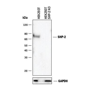
Western blot shows lysates of HEK293T human embryonic kidney parental cell line and SHP-2 knockout HEK293T cell line (KO). PVDF membrane was probed with 2 µg/mL of Rat Anti-Human/Mouse/Rat SHP-2 Monoclonal Antibody (Catalog # MAB1894) followed by HRP-conjugated Anti-Rat IgG Secondary Antibody (Catalog # HAF005). A specific band was detected for SHP-2 at approximately 80 kDa (as indicated) in the parental HEK293T cell line, but is not detectable in knockout HEK293T cell line. GAPDH (Catalog # MAB5718) is shown as a loading control. This experiment was conducted under reducing conditions and using Immunoblot Buffer Group 1.Applications for Human/Mouse/Rat SHP-2 Antibody
Knockout Validated
Western Blot
Sample: HepG2 human hepatocellular carcinoma cell line, TS1 mouse helper T cell line, and PC-12 rat adrenal pheochromocytoma cell line
Formulation, Preparation, and Storage
Purification
Reconstitution
Formulation
Shipping
Stability & Storage
- 12 months from date of receipt, -20 to -70 °C as supplied.
- 1 month, 2 to 8 °C under sterile conditions after reconstitution.
- 6 months, -20 to -70 °C under sterile conditions after reconstitution.
Background: SHP-2
Src-Homology domain-2 containing protein tyrosine Phosphatase 2 (SHP-2), also called protein tyrosine phosphatase, non-receptor type 11 (PTPN11), PTP1D, PTP2C, and SYP, is an enzyme that dephosphorylates tyrosine residues in proteins. The protein contains two Src homology 2 (SH2) domains, which both regulate the activity of the enzyme (1) and allow it to selectively bind to SH2 sites on proteins such as Dok1, IRS1, and the insulin receptor (2). SHP-2 plays a unique stimulatory role in cell signaling. Cells lacking SHP-2 have poor mobility because the hyper-phosphorylation of FAK and other proteins in the focal adhesion complex (3) prevents turnover of cellular attachment points. Without SHP-2, sustained ERK stimulation does not take place (4). The Y992 phosphorylation site of EGFR is a particularly good substrate for SHP-2 (5).
References
- Zhao, Z. et al. (1994) J. Biol. Chem. 269:8780.
- Clemmons, D.R. and Maile, L.A. (2005) Mol. Endocrinol. 19:1.
- von Wichert, G. et al. (2003) EMBO J. 22:5023.
- Maroun, C.R. et al. (2000) Mol. Cell. Biol. 20:8513.
- Sugimoto, S. et al. (1993) J. Biol. Chem. 269:22771.
Long Name
Alternate Names
Gene Symbol
UniProt
Additional SHP-2 Products
Product Documents for Human/Mouse/Rat SHP-2 Antibody
Product Specific Notices for Human/Mouse/Rat SHP-2 Antibody
For research use only

