Mouse TGF-beta RII Antibody
R&D Systems, part of Bio-Techne | Catalog # AF532

Key Product Details
Species Reactivity
Validated:
Cited:
Applications
Validated:
Cited:
Label
Antibody Source
Product Specifications
Immunogen
Ile24-Asp184
Accession # Q62312
Specificity
Clonality
Host
Isotype
Scientific Data Images for Mouse TGF-beta RII Antibody
Detection of Mouse TGF-beta RII by Western Blot.
Western blot shows lysates of mouse brain tissue. PVDF membrane was probed with 2 µg/mL of Goat Anti-Mouse TGF-beta RII Antigen Affinity-purified Polyclonal Antibody (Catalog # AF532) followed by HRP-conjugated Anti-Goat IgG Secondary Antibody (HAF019). A specific band was detected for TGF-beta RII at approximately 75 kDa (as indicated). This experiment was conducted under reducing conditions and using Immunoblot Buffer Group 8.Detection of TGF-beta RII in Mouse splenocytes by Flow Cytometry
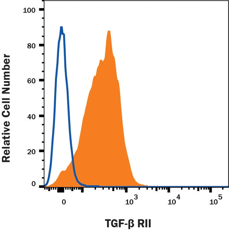
Mouse splenocytes were stained with Goat Anti-Mouse TGF-beta RII Antigen Affinity-purified Polyclonal Antibody (Catalog # AF532, filled histogram) or isotype control antibody (Catalog # AB-108-C, open histogram) followed by Phycoerythrin-conjugated Anti-Goat IgG Secondary Antibody (Catalog # F0107). View our protocol for Staining Membrane-associated Proteins.Applications for Mouse TGF-beta RII Antibody
CyTOF-ready
Flow Cytometry
Sample: Mouse splenocytes
Western Blot
Sample: Mouse brain tissue
Reviewed Applications
Read 1 review rated 5 using AF532 in the following applications:
Formulation, Preparation, and Storage
Purification
Reconstitution
Formulation
*Small pack size (-SP) is supplied either lyophilized or as a 0.2 µm filtered solution in PBS.
Shipping
Stability & Storage
- 12 months from date of receipt, -20 to -70 °C as supplied.
- 1 month, 2 to 8 °C under sterile conditions after reconstitution.
- 6 months, -20 to -70 °C under sterile conditions after reconstitution.
Background: TGF-beta RII
Most cell types express three sizes of receptors for TGF-beta. These are designated Type I (53 kDa), Type II (70 - 85 kDa), and Type III (250 - 350 kDa). The Type III receptor, a proteoglycan that exists in membrane-bound and soluble forms, binds TGF-beta 1, TGF-beta 2, and TGF-beta 3 but does not appear to be involved in signal transduction. The Type II receptor is a membrane-bound serine/threonine kinase that binds TGF-beta 1 and TGF-beta 3 with high affinity and TGF-beta 2 with a much lower affinity. The Type I receptor is also a membrane-bound serine/threonine kinase that apparently requires the presence of the Type II receptor to bind TGF-beta. Current evidence suggests that signal transduction requires the cytoplasmic domains of both the Type I and Type II receptors.
The recombinant soluble TGF-beta Type II receptor is capable of binding TGF-beta 1, TGF-beta 3, and TGF-beta 5 with sufficient affinity to act as an inhibitor of these isoforms at high concentrations. The soluble receptor also binds TGF-beta 2, but with an affinity at least two orders of magnitude lower. Binding of TGF-beta 1, TGF-beta 3, and TGF-beta 5 to the soluble TGF-beta Type II receptor can also be demonstrated by using the soluble receptor as a capture agent on ELISA plates and this observation has been used as the basis for the development of immunoassays for these isoforms of TGF-beta.
References
- Miyazono, K. et al. (1994) Adv. in Immunol. 55:181.
Long Name
Alternate Names
Gene Symbol
UniProt
Additional TGF-beta RII Products
Product Documents for Mouse TGF-beta RII Antibody
Product Specific Notices for Mouse TGF-beta RII Antibody
For research use only

