Mouse TIM-1/KIM-1/HAVCR Antibody Best Seller
R&D Systems, part of Bio-Techne | Catalog # AF1817


Key Product Details
Species Reactivity
Validated:
Cited:
Applications
Validated:
Cited:
Label
Antibody Source
Product Specifications
Immunogen
Tyr22-Thr212
Accession # NP_001160104
Specificity
Clonality
Host
Isotype
Scientific Data Images for Mouse TIM-1/KIM-1/HAVCR Antibody
Detection of Mouse TIM-1/KIM-1/HAVCR by Western Blot.
Western blot shows lysates of NIH-3T3 mouse embryonic fibroblast cell line. PVDF membrane was probed with 0.25 µg/mL of Goat Anti-Mouse TIM-1/KIM-1/HAVCR Antigen Affinity-purified Polyclonal Antibody (Catalog # AF1817) followed by HRP-conjugated Anti-Goat IgG Secondary Antibody (Catalog # HAF019). A specific band was detected for TIM-1/KIM-1/HAVCR at approximately 70-80 kDa (as indicated). This experiment was conducted under reducing conditions and using Immunoblot Buffer Group 1.Detection of Mouse TIM-1/KIM-1/HAVCR by Western Blot
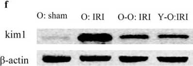
Exogenous biological renal support improved renal IRI and decreased mortality and serum Cr, BUN levels in old IRI mice. (A) Survival curves for the old IRI mice at 72 hours. (B) Cr levels in the old mice. (C) BUN levels in the old mice. (D) Representative photographs of kidney sections from the old mice stained with periodic acid–Schiff (400× magnification). (E) Renal tubular injury score. (F) The levels of Kim1 in kidney extracts from the old mice, as measured by western blotting. Gels were performed under the same experimental conditions. (G) Quantitative analyses of the band densities of Kim1 expression. Values are presented as means ± SDs. ▲P < 0.05, ▲▲P < 0.01 vs. O: sham; *P < 0.05, **P < 0.01 vs. O: IRI. BUN, blood urea nitrogen Cr, serum creatinine; SD, standard deviation. Image collected and cropped by CiteAb from the following publication (https://pubmed.ncbi.nlm.nih.gov/30978173), licensed under a CC-BY license. Not internally tested by R&D Systems.Applications for Mouse TIM-1/KIM-1/HAVCR Antibody
Western Blot
Sample: NIH‑3T3 mouse embryonic fibroblast cell line
Reviewed Applications
Read 1 review rated 5 using AF1817 in the following applications:
Formulation, Preparation, and Storage
Purification
Reconstitution
Formulation
Shipping
Stability & Storage
- 12 months from date of receipt, -20 to -70 °C as supplied.
- 1 month, 2 to 8 °C under sterile conditions after reconstitution.
- 6 months, -20 to -70 °C under sterile conditions after reconstitution.
Background: TIM-1/KIM-1/HAVCR
TIM-1 (T cell-immunoglobulin-mucin; also KIM-1 and HAVCR) is a 70-80 kDa, type I transmembrane glycoprotein member of the TIM family of immunoglobulin superfamily molecules (1-4). This gene family is involved in the regulation of Th1 and Th2-cell-mediated immunity. In mouse, there are eight known TIM genes (# 1-8) vs. only three genes in human (# 1, 3, and 4) (1, 2). Mouse TIM-1 and -2 are counterparts of human TIM-1 while mouse TIM-5 through 8 have no human counterparts (2). Mouse TIM-1 is synthesized as a 305 amino acid (aa) precursor that contains a 21 aa signal sequence, a 216 aa extracellular domain (ECD), a 21 aa transmembrane segment and a 47 aa cytoplasmic domain (5, 6). The ECD contains one V-type Ig-like domain and a mucin region characterized by multiple T-S-P motifs. The mucin region undergoes extensive O-linked glycosylation. The mouse TIM-1 gene is highly polymorphic and, based on rat, may undergo alternate splicing (4, 6). For instance, HBA mice show a 15 aa deletion in the mucin region that occurs in BALB/c mice (6). This difference is associated with a decreased susceptibility to asthma. Other polymorphisms are also documented (6). In human, TIM-1 is known to circulate as a soluble form. It undergoes constitutive cleavage by an undefined MMP, releasing a 75-85 kDa soluble molecule (5). The same thing might be expected in mouse. The ECD of mouse TIM-1 is 50%, 39%, and 80% aa identical to human, canine and rat TIM-1 ECD, respectively. The only two reported ligands for TIM-1 are TIM-4 and the hepatitis A virus (8, 9). However, others are believed to exist, and based on the ligand for TIM-3, one possibility might be an S-type lectin (10). TIM-1 ligation induces T cell proliferation and promotes cytokine production (1, 10). In particular, it induces IL-4 production, and requires the cytoplasmic tyrosine phosphorylation motif (5).
References
- Meyers, J.H. et al. (2005) Trends Mol. Med. 11:1471.
- Kuchroo, V.K. et al. (2003) Nat. Rev. Immunol. 3:454.
- Mariat, C. et al. (2005) Phil. Trans. R. Soc. B 360:1681.
- Ichimura, T. et al. (1998) J. Biol. Chem. 273:4135.
- de Souza, A.J. et al. (2005) Proc. Natl. Acad. Sci. USA 102:17113.
- McIntire, J.J. et al. (2001) Nat. Immunol. 2:1109.
- Bailly, V. et al. (2002) J. Biol. Chem. 277:39739.
- Feigelstock, D. et al. (1998) J. Virol. 72:6621.
- Zhu, C. et al. (2005) Nat. Immunol. 6:1245.
- Meyers, J.H. et al. (2005) Nat. Immunol. 6:455.
Long Name
Alternate Names
Gene Symbol
UniProt
Additional TIM-1/KIM-1/HAVCR Products
Product Documents for Mouse TIM-1/KIM-1/HAVCR Antibody
Product Specific Notices for Mouse TIM-1/KIM-1/HAVCR Antibody
For research use only
