Human xCT/SLC7A11 Antibody
R&D Systems, part of Bio-Techne | Catalog # MAB11251

Key Product Details
Species Reactivity
Human
Applications
Flow Cytometry, Western Blot
Label
Unconjugated
Antibody Source
Monoclonal Mouse IgG1 Clone # 1057408
Product Specifications
Immunogen
Synthetic peptide within amino acids 1-50 of the Human xCT/SLC7A11 protein
Met1-Leu501
Accession # Q9UPY5
Met1-Leu501
Accession # Q9UPY5
Clonality
Monoclonal
Host
Mouse
Isotype
IgG1
Scientific Data Images for Human xCT/SLC7A11 Antibody
Detection of Human xCT/SLC7A11 by Western Blot.
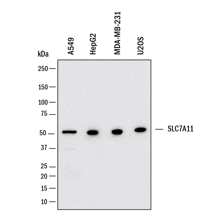
Western blot shows lysates of A549 human lung carcinoma cell line, HepG2 human hepatocellular carcinoma cell line, MDA-MB-231 human breast cancer cell line, U2OS human osteosarcoma cell line. PVDF membrane was probed with 2 µg/mL of Mouse Anti-Human xCT/SLC7A11 Monoclonal Antibody (Catalog # MAB11251) followed by HRP-conjugated Anti-Mouse IgG Secondary Antibody (Catalog # HAF018). A specific band was detected for xCT/SLC7A11 at approximately 55 kDa (as indicated). This experiment was conducted under reducing conditions and using Western Blot Buffer Group 1.Detection of xCT/SLC7A11 in HEK293 Human Cell Line Transfected with Human xCT/SLC7A11 and eGFP by Flow Cytometry.
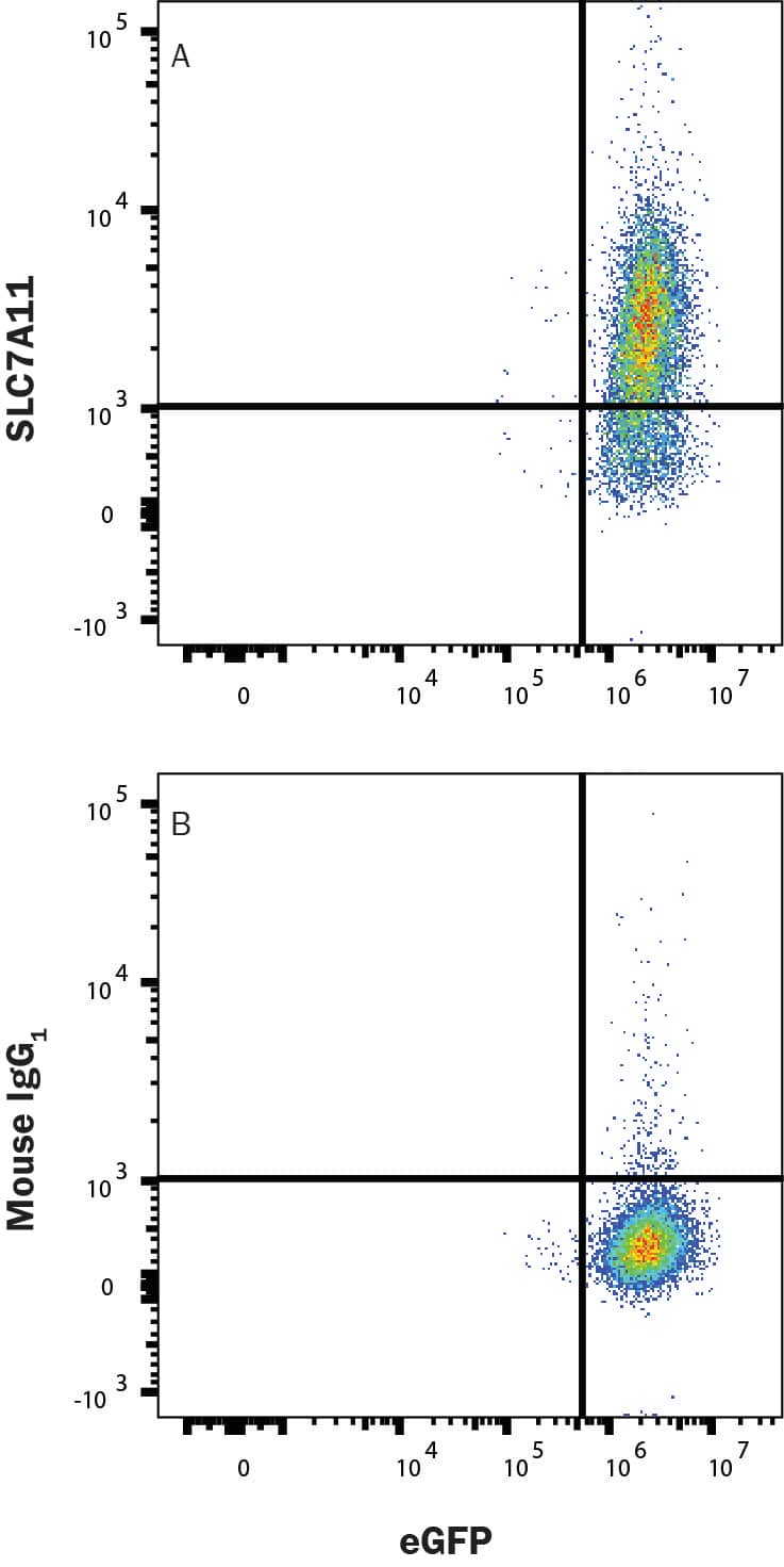
HEK293 human embryonic kidney cell line transfected with human xCT/SLC7A11 and eGFP was stained with (A) Mouse Anti-Human xCT/SLC7A11 Monoclonal Antibody (Catalog # MAB11251) or (B) Mouse IgG1 control antibody staining (MAB002) followed by Allophycocyanin-conjugated Anti-Mouse IgG Secondary Antibody (F0101B). View our protocol for Staining Membrane-associated Proteins.Applications for Human xCT/SLC7A11 Antibody
Application
Recommended Usage
Flow Cytometry
0.25 µg/mL
Sample: HEK293 human embryonic kidney cell line transfected with human xCT/SLC7A11 and eGFP
Sample: HEK293 human embryonic kidney cell line transfected with human xCT/SLC7A11 and eGFP
Western Blot
2 µg/mL
Sample: Lysates of A549 human lung carcinoma cell line, HepG2 human hepatocellular carcinoma cell line, MDA-MB-231 human breast cancer cell line, U2OS human osteosarcoma cell line.
Sample: Lysates of A549 human lung carcinoma cell line, HepG2 human hepatocellular carcinoma cell line, MDA-MB-231 human breast cancer cell line, U2OS human osteosarcoma cell line.
Formulation, Preparation, and Storage
Purification
Protein A or G purified from hybridoma culture supernatant
Reconstitution
Reconstitute at 0.5 mg/mL in sterile PBS. For liquid material, refer to CoA for concentration.
Formulation
Lyophilized from a 0.2 μm filtered solution in PBS with Trehalose *Small pack size (SP) is supplied either lyophilized or as a 0.2 µm filtered solution in PBS.
Shipping
Lyophilized product is shipped at ambient temperature. Liquid small pack size (-SP) is shipped with polar packs. Upon receipt, store immediately at the temperature recommended below.
Stability & Storage
Use a manual defrost freezer and avoid repeated freeze-thaw cycles.
- 12 months from date of receipt, -20 to -70 °C as supplied.
- 1 month, 2 to 8 °C under sterile conditions after reconstitution.
- 6 months, -20 to -70 °C under sterile conditions after reconstitution.
Background: xCT/SLC7A11
References
- Liu, J. Xia, X. & Huang, P. (2020). xCT: A Critical Molecule That Links Cancer Metabolism to Redox Signaling. Molecular therapy : the journal of the American Society of Gene Therapy. https://doi.org/10.1016/j.ymthe.2020.08.021.
- Koppula, P. Zhang, Y. Zhuang, L. & Gan, B. (2018). Amino acid transporter SLC7A11/xCT at the crossroads of regulating redox homeostasis and nutrient dependency of cancer. Cancer communications. https://doi.org/10.1186/s40880-018-0288-x.
- Lin, W. Wang, C., Liu, G., Bi, C., Wang, X. Zhou, Q., & Jin, H. (2020). SLC7A11/xCT in cancer: biological functions and therapeutic implications. American journal of cancer research.
- xCT: Uniprot (Q9UPY5).
- Koppula, P. Zhuang, L. & Gan, B. (2020). Cystine transporter SLC7A11/xCT in cancer: ferroptosis, nutrient dependency, and cancer therapy. Protein & cell. https://doi.org/10.1007/s13238-020-00789-5.
- Liu, L. Liu, R. Liu, Y. Li, G., Chen, Q. Liu, X., & Ma, S. (2020). Cystine-glutamate antiporter xCT as a therapeutic target for cancer. Cell biochemistry and function. https://doi.org/10.1002/cbf.3581.
- Cui, Q. Wang, J. Q. Assaraf, Y. G. Ren, L. Gupta, P. Wei, L., Ashby, C. R. Jr. Yang, D. H. & Chen, Z. S. (2018). Modulating ROS to overcome multidrug resistance in cancer. Drug resistance updates : reviews and commentaries in antimicrobial and anticancer chemotherapy. https://doi.org/10.1016/j.drup.2018.11.001.
Long Name
Cationic 1/Solute Carrier Family 7 Member 11
Alternate Names
CCBR1, SLC7A11
Gene Symbol
SLC7A11
UniProt
Additional xCT/SLC7A11 Products
Product Documents for Human xCT/SLC7A11 Antibody
Product Specific Notices for Human xCT/SLC7A11 Antibody
For research use only
Loading...
Loading...
Loading...
Loading...
Loading...

