Products | Resources | Background Information
Changes in cell membrane potential are generated by the movement of ions across the cell membrane via ion channels and transporter proteins. These voltage changes occur in response to external stimuli, such as neurotransmitter release, and can be measured from single ion channels and cells to whole organs. This electrical activity of cells is important for the normal functioning of organisms and has been found to be altered in certain disease states.
Products for Electrophysiology
GPCR Ligands
GPCR Ligands
G-protein coupled receptors (GPCRs) are membrane proteins that mediate downstream signaling of many peptides, hormones and neurotransmitters. Compounds acting at GPCRs enable manipulation of signaling pathways and associated biological responses.
Search for your target in the search bar above or Browse pharmacology groups on tocris.com by clicking below
Neurotransmitter Transporter Inhibitors
Neurotransmitter Transporter Inhibitors
Neurotransmitter transporters carry neurotransmitters across biological membranes, and are found in high concentrations in synaptic clefts where they terminate the effect of a neurotransmitter. They often rely on an electrochemical gradient across the membrane to provide energy for transport.
Fluorescence Imaging
Fluorescence Imaging
Fluorescent dyes and probes enable imaging of proteins and cellular structures in live and fixed cells. Our range includes microtubule probes, cell membrane stains, and neuron and astrocyte probes.
Caged Compounds
Caged Compounds
Caged compounds are small molecules that have been chemically protected with a photolabile group, which renders the compound biologically inactive. Activation with light removes the protective group, releasing the active compound adjacent to its target.
Chemogenetics
Chemogenetics
Chemogenetic compounds enable the control of excitable cell activity via small molecules that selectively target genetically modified GPCRs (DREADDs and DREADD ligands) or engineered chimeric ion channels (PSAMs and PSEMs). DREADD ligands can also be used to investigate signaling pathways downstream of GPCR activation.
Ion Channel Modulators
Ion Channel Modulators
Ion channels are classified based on their gating mechanism and their ion selectivity. The distribution of ions across a cell membrane determines the membrane potential, and small molecule and peptide ion channel modulators, including toxins, are commonly used in electrophysiology to manipulate ion flow. View full product ranges at tocris.com
Image Synaptic Activity with FFNs
False Fluorescent Neurotransmitters (FFNs) are fluorescent substrates for neurotransmitter transporters such as VMAT2 and NET. They label neurons and synaptic vesicles expressing a transporter and enable imaging and tracking of neurotransmitter release and exocytotic events from synaptic vesicles in cultured neurons and in vivo.
FFN 200 (Cat. No. 5911) used to image vesicle clusters in the axons of cultured dopaminerigc neurons, and exocytosis of vesicles on high potassium stimulation (time lapse at 12 images/min).
Electrophysiology Resources
Chemogenetics Research Bulletin
Chemogenetics Research Bulletin
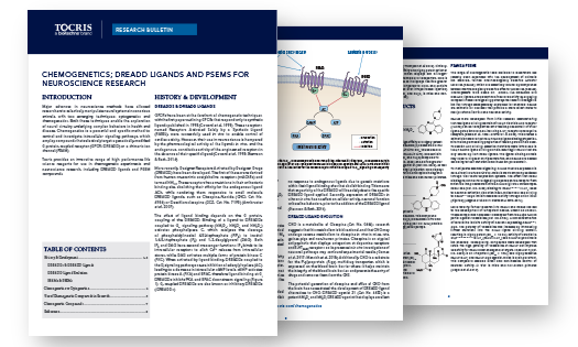
Produced by our in-house experts, the Chemogenetics Research Bulletin provides an introduction to chemogenetics methods to manipulate neuronal activity in vitro and in vivo. The development of genetically modified receptors and ion channels, including RASSLs, DREADDs and PSAMs, as well as the use of chemogenetic compounds is discussed.
Fluorescent Probes and Dyes Product Guide
Fluorescent Probes and Dyes Product Guide
Our Fluorescent Probes and Dyes Research Guide provides a background to the use of Fluorescent Probes and Dyes, as well as a comprehensive list of our product range which includes:
- Fluorescent Dyes, including dyes for Flow Cytometry
- Fluorescent Probes
- Bioluminescent Substrates
- Fluorogenic Dyes for Light-Up Aptamers
- Fluorescent Probes for Imaging Bacteria
- TSA Reagents for Enhancing IHC, ICC & FISH Signals
Related Research Products for Electrophysiology
RNAscope® ISH Technology
RNAscope® ISH Technology
Visualize Multiple Markers Simultaneously in Tissue.
Detect, characterize and localize mRNA in the nervous system with RNAscope ISH.
Cell Culture Reagents
Cell Culture Reagents
Bio-Techne offers a comprehensive portfolio of reagents for cell culture including classic media, balanced salt solutions, supplements, and basement membrane extracts.
GPCR Antibodies
GPCR Antibodies
Despite being the largest family of integral membrane proteins and having a key role in signal transduction, developing antibodies to detect GPCRs is notoriously difficult. Trust the industry-leading expertise of R&D Systems for your GPCR antibodies.
Broadly, electrophysiology is the measurement of electrical current across a cell membrane, in a tissue or in a whole organ. As a biomedical research technique, electrophysiology is most widely used in neuroscience and cardiovascular research to investigate the electrical properties of central or peripheral neurons, and cardiomyocytes, respectively. However, electrophysiology methods can also be used in microbiology to study ion channels in bacteria, or in endocrinology to study the excitable properties of pancreatic β-cells, for example.
Electrophysiology techniques are used in multiple experimental situations, depending on the investigation being performed. In vitro, patch clamping can be used to study isolated sections of membrane, specific ion channels or receptors endogenously or artificially expressed in cells, ex vivo whole cells, or cell cultures derived from stem cells. Additionally, in neuroscience research brain slices maintained in artificial cerebrospinal fluid (aCSF) can also be used. In vivo, electrophysiological techniques can be used to measure local field potentials, either with single electrodes or multi-electrode arrays, or to investigate the activity of individual neurons by whole-cell patch clamping.
Small molecules are utilized in all types of electrophysiology experiments, for example to manipulate and investigate the function of an individual ion channel or receptor; to explore the physiological consequences of transporter or enzyme inhibition at a molecular, cellular or systems level; and to alter the activity of a single cell or population of cells, and investigate the connectivity of those cells with other areas of the brain.
‘Patch Clamping’ is an electrophysiology technique that involves intracellular recording . This technique, developed by Bert Sakmann and Erwin Neher in the late 1970s, is used to study changes in ion flow (ionic currents) in an isolated cell or ‘patch’ of cell membrane. There are two different patch clamp methods: the voltage clamp technique where the voltage across a membrane is controlled and the resulting current changes are recorded; and the current clamp technique, in which the current passing across a membrane is controlled and changes in voltage are measured. In neurons, the change in voltage is in the form of action potentials.
In patch clamp experiments a micropipette filled with an electrolyte solution, and a recording pipette are brought into contact with the cell membrane. The electrolyte solution used depends on the type of recording being conducted, and the exact experimental set up. Most patch clamping is done in vitro, however recent advances in the technique have allowed researchers to perform whole-cell patch clamping in vivo, in either sedated or awake, head-fixed rodents. Additionally, advances in robotics and automation have enabled patch clamp methods to be developed for high throughput screening experiments.
Types of Patch Clamp
- Cell-attached patch where the micropipette is sealed to the cell membrane, but the membrane remains intact and the cell is not punctured. This allows recording of ion currents through channels located in the ‘patch’ of membrane at the end of the micropipette. Small molecules can be included in the pipette electrolyte solution to modulate the function of ion channels or GPCRs being studied.
- Inside-out patch where the patch of membrane attached to the micropipette is detached from the rest of the cell. This allows manipulation of the environment at the intracellular surface of the membrane.
- Whole-cell patch where suction is applied through the micropipette following attachment to the cell, to pierce the cell membrane allowing the interior of the pipette to access the interior of the cell. This allows recording of currents through multiple channels simultaneously.
- Outside-out patch where, following set up of a whole-cell patch, the micropipette is pulled away from the cells, pulling sections of the membrane with it. The membrane sections detach from the rest of the cell and their ends attached together to form a convex membrane section on the end of the pipette. The original outer face of the cell membrane would now face outward from the electrode. This allows small molecules to be applied to the external face of the membrane via the experimental bath solution, or in the internal surface via the electrode solution.
Measuring the activity of individual neurons in vivo, also known as single-unit recording, can be achieved by using a microelectrode with a tip size of approximately 1 µm. Action potentials recorded in this way display a similar profile to those recorded by intracellular electrophysiology methods, albeit with a smaller signal. Single unit recordings allow for greater spatial and temporal resolution when accessing the relationship between neuronal activity, brain structure and function, and behaviors. DREADDs (Designer Receptors Exclusively Activated by Designer Drugs) and DREADD ligands are widely used tools for manipulating neuronal activity in vivo. DREADDS are genetically modified GPCRs that are unresponsive to endogenous compounds. When expressed in cells they can be manipulated using DREADD ligands allowing precise control and investigation of neuronal signaling.
In vivo electrophysiology techniques also enable the recording of extracellular local field potentials (LFPs), which are transient electrical signals generated by the synchronous electrical activity of individual cells in the tissue being investigated. They are recorded by electrodes placed near the generating cells, and the signal measured reflects the sum of action potentials from multiple cells approximately 50 - 350 µm from the tip of the electrode. This form of electrophysiology is often called multi-unit recording.
Recent advances in experimental hardware for in vivo electrophysiology in neuroscience allow researchers to measure LFPs in multiple areas of the brain, or at multiple sites within a specific brain area, at the same time. These advances include recording electrodes with multiple recording sites spaced a regular intervals on their shank, and multi-electrode arrays (MEAs) that can be chronically implanted to allow long-term measurements of neuronal activity. MEAs allow researchers to investigate how the activity in a brain area may be associated with physiological and behavioral outcomes in animal models.


