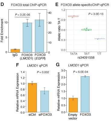
Chromatin Immunoprecipitation (ChIP): FOXO3 Antibody [NBP2-16521] - (D) ChIP assay for LMOD1 and EGFR (as a positive control region) in HCASMC chromatin lysates immunoprecipitated with antibodies to FOXO3 or a negative control rabbit IgG. n = 3. (E) Allele specific ChIP (haploChIP) for FOXO3 protein in DNA derived from cultured HCASMC ChIP experiments. DNA from cell line homozygous for the ancestral allele was used as a positive control and arbitrarily set to 1. Values represent mean +/- standard deviation of triplicates. Similar results were observed from n = 4 independent lines for each genotype. (F) Quantitative RT-PCR analysis of LMOD1 in HCASMCs following knock down of endogenous FOXO3 (F) or overexpression of FOXO3 (G). The results were reproduced in n = 3 independent experiments. Image collected and cropped by CiteAb from the following publication (https://dx.plos.org/10.1371/journal.pgen.1007755) licensed under a CC-BY license.
Western Blot: FOXO3 Antibody [NBP2-16521]
Western Blot: FOXO3 Antibody [NBP2-16521] - 50 ug of mouse brain whole cell lysate). Antibody at 1:500.
Western Blot: FOXO3 Antibody [NBP2-16521]
Western Blot: FOXO3 Antibody [NBP2-16521] - Non-transfected (-) and transfected (+) C2C12 whole cell extracts (30 ug) were separated by 7.5% SDS-PAGE, and the membrane was blotted with FOXO3A antibody [C3], C-term.
Immunohistochemistry-Paraffin: FOXO3 Antibody [NBP2-16521]
Immunohistochemistry-Paraffin: FOXO3 Antibody [NBP2-16521] - Huh-7 xenograft, using FOXO3A antibody at 1:500 dilution. Antigen Retrieval: Trilogy™ (EDTA based, pH 8.0) buffer, 15min.
Immunohistochemistry-Paraffin: FOXO3 Antibody [NBP2-16521]
Immunohistochemistry-Paraffin: FOXO3 Antibody [NBP2-16521] - Rat colon. FOXO3A stained by FOXO3A antibody [C3], C-term diluted at 1:2000.Antigen Retrieval: Citrate buffer, pH 6.0, 15 min.
Immunohistochemistry-Paraffin: FOXO3 Antibody [NBP2-16521]
Immunohistochemistry-Paraffin: FOXO3 Antibody [NBP2-16521] - Mouse testis. FOXO3A stained by FOXO3A antibody [C3], C-term diluted at 1:2000.Antigen Retrieval: Citrate buffer, pH 6.0, 15 min.
Immunohistochemistry-Paraffin: FOXO3 Antibody [NBP2-16521]
Immunohistochemistry-Paraffin: FOXO3 Antibody [NBP2-16521] - Rat brain. FOXO3A stained by FOXO3A antibody [C3], C-term diluted at 1:2000.Antigen Retrieval: Citrate buffer, pH 6.0, 15 min.
Immunohistochemistry-Paraffin: FOXO3 Antibody [NBP2-16521]
Immunohistochemistry-Paraffin: FOXO3 Antibody [NBP2-16521] - Mouse brain. FOXO3A stained by FOXO3A antibody [C3], C-term diluted at 1:2000. Antigen Retrieval: Citrate buffer, pH 6.0, 15 minutes.
Immunoprecipitation: FOXO3 Antibody [NBP2-16521]
Immunoprecipitation: FOXO3 Antibody [NBP2-16521] - Jurkat whole cell lysate/extract A. Control with 2 ug of preimmune rabbit IgG B. Immunoprecipitation of FOXO3A protein by 2 ug of FOXO3A antibody 5% SDS-PAGE The immunoprecipitated FOXO3A protein was detected by FOXO3A antibody diluted at 1:1000. EasyBlot anti-rabbit IgG was used as a secondary reagent.
Western Blot: FOXO3 Antibody [NBP2-16521] -
Western Blot: FOXO3 Antibody [NBP2-16521] - LMOD1 expression is mediated by PDGF-BB-FOXO3 signaling cascade.(A) UCSC Browser screenshot of the LMOD1 CAD locus at chromosome 1q32.1 highlighting the candidate causal variant, rs34091558, overlapping ATAC-seq open chromatin & RNA-seq tracks in HCASMCs treated with PDGF-BB (n = 2 biological replicates). Genomic coordinates refer to hg19 assembly. Quantitative RT-PCR (B) & Western blotting (C) data revealing PDGF-BB & phosphorylated-FOXO3 (P-FOXO3) mediated LMOD1 expression. (D) Co-expression microarray analysis performed in a cohort of carotid atherosclerotic plaques (n = 127) indicating that LMOD1 & FOXO3 are positively correlated in arterial tissues. Image collected & cropped by CiteAb from the following publication (https://pubmed.ncbi.nlm.nih.gov/30444878), licensed under a CC-BY license. Not internally tested by Novus Biologicals.
Immunoprecipitation: FOXO3 Antibody [NBP2-16521] -
Immunoprecipitation: FOXO3 Antibody [NBP2-16521] - Chemotherapy-induced A2BR expression promotes FOXO3 binding on pluripotency factor genes through decreased H3K27me3 & increased H3K27ac chromatin marks. A & B, MDA-MB-231 NTC or A2BR knockdown subclones were transfected with pLX304 (empty vector, EV) or pLX304 encoding A2BR. Cells were treated with vehicle (V) or 10 nM paclitaxel (P) for 72 h & chromatin immunoprecipitation (ChIP) was performed with antibody against FOXO3. Primers flanking the FOXO3 binding site in the NANOG, SOX2 & KLF4 gene (A) were used for qPCR (B; mean ± SEM; n = 3); **p < 0.01, ***p < 0.001 vs. NTC/EV-V; #p < 0.05, ##p < 0.01 vs. NTC/EV-P; ^^p < 0.01, ^^^p < 0.0001 vs. A2BR shRNA/EV-P; ns, not significant. C & E, MDA-MB-231 NTC or A2BR knockdown subclones were treated with V or P for 72 h & immunoblot assays were performed. D, MDA-MB-231 NTC or A2BR knockdown subclones were treated with V or P for 72 h. Cytosolic & nuclear lysates were prepared, & immunoblot assays were performed. F-H, MDA-MB-231 NTC or A2BR knockdown subclones were treated with V or 10 nM P for 72 h. ChIP was performed using antibodies against H3K27me3 (F), H3K27ac (G), or Histone H3 (H) followed by qPCR with primers flanking FOXO3 binding sites in the NANOG, SOX2 & KLF4 gene (mean ± SEM; n = 3); *p < 0.05, **p < 0.01, ***p < 0.001 vs. NTC-V; #p < 0.05, ##p < 0.01, ###p < 0.001 vs. NTC-P; ns, not significant. Image collected & cropped by CiteAb from the following publication (https://pubmed.ncbi.nlm.nih.gov/35401817), licensed under a CC-BY license. Not internally tested by Novus Biologicals.
Immunoprecipitation: FOXO3 Antibody [NBP2-16521] -
Immunoprecipitation: FOXO3 Antibody [NBP2-16521] - SMARCD3 knockdown blocks paclitaxel-induced FOXO3 binding on pluripotency factor genes & inhibits BCSC enrichment. A, MDA-MB-231 cells were transfected with vector encoding NTC or either of two shRNAs targeting SMARCD3 (#1 & #2), & immunoblot assay was performed. B-D, MDA-MB-231 NTC or SMARCD3 knockdown subclones were treated with vehicle (V) or 10 nM paclitaxel (P) for 72 h, & ALDH (B; mean ± SEM; n = 3), mammosphere (C; mean ± SEM; n = 4), & qPCR (D; mean ± SEM; n = 3) assays were performed; *p < 0.05, **p < 0.01, ***p < 0.001 vs. NTC-V; #p < 0.05, ##p < 0.01, ###p < 0.001 vs. NTC-P; ns, not significant. E-I, MDA-MB-231 NTC or SMARCD3 knockdown subclones were treated with V or 10 nM P for 72 h. ChIP was performed using antibodies against FOXO3 (E), H3K27me3 (F), H3K27ac (G), KDM6A (H), or p300 (I), followed by qPCR with primers flanking FOXO3 binding sites in the NANOG, SOX2 & KLF4 gene (mean ± SEM; n = 4); *p < 0.05, **p < 0.01, ***p < 0.001 vs. NTC-V; ##p < 0.01, ###p < 0.001 vs. NTC-P; ns, not significant. Image collected & cropped by CiteAb from the following publication (https://pubmed.ncbi.nlm.nih.gov/35401817), licensed under a CC-BY license. Not internally tested by Novus Biologicals.
Immunoprecipitation: FOXO3 Antibody [NBP2-16521] -
Immunoprecipitation: FOXO3 Antibody [NBP2-16521] - Chemotherapy-induced A2BR expression promotes FOXO3 binding on pluripotency factor genes through decreased H3K27me3 & increased H3K27ac chromatin marks. A & B, MDA-MB-231 NTC or A2BR knockdown subclones were transfected with pLX304 (empty vector, EV) or pLX304 encoding A2BR. Cells were treated with vehicle (V) or 10 nM paclitaxel (P) for 72 h & chromatin immunoprecipitation (ChIP) was performed with antibody against FOXO3. Primers flanking the FOXO3 binding site in the NANOG, SOX2 & KLF4 gene (A) were used for qPCR (B; mean ± SEM; n = 3); **p < 0.01, ***p < 0.001 vs. NTC/EV-V; #p < 0.05, ##p < 0.01 vs. NTC/EV-P; ^^p < 0.01, ^^^p < 0.0001 vs. A2BR shRNA/EV-P; ns, not significant. C & E, MDA-MB-231 NTC or A2BR knockdown subclones were treated with V or P for 72 h & immunoblot assays were performed. D, MDA-MB-231 NTC or A2BR knockdown subclones were treated with V or P for 72 h. Cytosolic & nuclear lysates were prepared, & immunoblot assays were performed. F-H, MDA-MB-231 NTC or A2BR knockdown subclones were treated with V or 10 nM P for 72 h. ChIP was performed using antibodies against H3K27me3 (F), H3K27ac (G), or Histone H3 (H) followed by qPCR with primers flanking FOXO3 binding sites in the NANOG, SOX2 & KLF4 gene (mean ± SEM; n = 3); *p < 0.05, **p < 0.01, ***p < 0.001 vs. NTC-V; #p < 0.05, ##p < 0.01, ###p < 0.001 vs. NTC-P; ns, not significant. Image collected & cropped by CiteAb from the following publication (https://pubmed.ncbi.nlm.nih.gov/35401817), licensed under a CC-BY license. Not internally tested by Novus Biologicals.
Immunoprecipitation: FOXO3 Antibody [NBP2-16521] -
Immunoprecipitation: FOXO3 Antibody [NBP2-16521] - A2BR decreases H3K27me3 & increases H3K27ac marks through recruitment of KDM6A & p300 at FOXO3 binding sites of pluripotency factor genes. A & B, MDA-MB-231 NTC or A2BR knockdown subclones were transfected with pLX304 (empty vector, EV) or pLX304 encoding A2BR. Cells were treated with vehicle (V) or 10 nM paclitaxel (P) for 72 h. ChIP was performed using antibodies against KDM6A (A) or p300 (B) followed by qPCR with primers flanking FOXO3 binding sites in the NANOG, SOX2 & KLF4 gene (mean ± SEM; n = 3); *p < 0.05, **p < 0.01, ***p < 0.001 vs. NTC-V; ##p < 0.01, ###p < 0.001 vs. NTC-P; ^^p < 0.01, ^^^p < 0.0001 vs. A2BR shRNA/EV-P; ns, not significant. C, MDA-MB-231 NTC or A2BR knockdown subclones were treated with V or P for 72 h & immunoblot assays were performed. D, MDA-MB-231 NTC or A2BR knockdown subclones were treated with V or P for 72 h & nuclear lysates were prepared. Immunoprecipitation (IP) was performed using FOXO3 antibody or control IgG followed by immunoblot assays. NL, nuclear protein lysate. Image collected & cropped by CiteAb from the following publication (https://pubmed.ncbi.nlm.nih.gov/35401817), licensed under a CC-BY license. Not internally tested by Novus Biologicals.
Western Blot: FOXO3 Antibody [NBP2-16521] -
Western Blot: FOXO3 Antibody [NBP2-16521] - Various whole cell extracts (30 ug) were separated by 7.5% SDS-PAGE, and the membrane was blotted with FOXO3A antibody [C3], C-term diluted at 1:1000. The HRP-conjugated anti-rabbit IgG antibody was used to detect the primary antibody. Corresponding RNA expression data for the same cell lines are based on Human Protein Atlas program.

![Immunocytochemistry/ Immunofluorescence: FOXO3 Antibody [NBP2-16521] Immunocytochemistry/ Immunofluorescence: FOXO3 Antibody [NBP2-16521]](https://resources.bio-techne.com/images/products/FOXO3-Antibody-Immunocytochemistry-Immunofluorescence-NBP2-16521-img0023.jpg)
![Immunohistochemistry-Paraffin: FOXO3 Antibody [NBP2-16521] Immunohistochemistry-Paraffin: FOXO3 Antibody [NBP2-16521]](https://resources.bio-techne.com/images/products/FOXO3-Antibody-Immunohistochemistry-Paraffin-NBP2-16521-img0028.jpg)

![Western Blot: FOXO3 Antibody [NBP2-16521] Western Blot: FOXO3 Antibody [NBP2-16521]](https://resources.bio-techne.com/images/products/FOXO3-Antibody-Western-Blot-NBP2-16521-img0021.jpg)
![Western Blot: FOXO3 Antibody [NBP2-16521] Western Blot: FOXO3 Antibody [NBP2-16521]](https://resources.bio-techne.com/images/products/FOXO3A-Antibody-Western-Blot-NBP2-16521-img0002.jpg)
![Western Blot: FOXO3 Antibody [NBP2-16521] Western Blot: FOXO3 Antibody [NBP2-16521]](https://resources.bio-techne.com/images/products/FOXO3-Antibody-Western-Blot-NBP2-16521-img0015.jpg)
![Immunohistochemistry-Paraffin: FOXO3 Antibody [NBP2-16521] Immunohistochemistry-Paraffin: FOXO3 Antibody [NBP2-16521]](https://resources.bio-techne.com/images/products/FOXO3A-Antibody-Immunohistochemistry-Paraffin-NBP2-16521-img0003.jpg)
![Immunohistochemistry-Paraffin: FOXO3 Antibody [NBP2-16521] Immunohistochemistry-Paraffin: FOXO3 Antibody [NBP2-16521]](https://resources.bio-techne.com/images/products/FOXO3-Antibody-Immunohistochemistry-Paraffin-NBP2-16521-img0024.jpg)
![Immunohistochemistry-Paraffin: FOXO3 Antibody [NBP2-16521] Immunohistochemistry-Paraffin: FOXO3 Antibody [NBP2-16521]](https://resources.bio-techne.com/images/products/FOXO3-Antibody-Immunohistochemistry-Paraffin-Negative-NBP2-16521-img0025.jpg)
![Immunohistochemistry-Paraffin: FOXO3 Antibody [NBP2-16521] Immunohistochemistry-Paraffin: FOXO3 Antibody [NBP2-16521]](https://resources.bio-techne.com/images/products/FOXO3-Antibody-Immunohistochemistry-Paraffin-NBP2-16521-img0026.jpg)
![Immunohistochemistry-Paraffin: FOXO3 Antibody [NBP2-16521] Immunohistochemistry-Paraffin: FOXO3 Antibody [NBP2-16521]](https://resources.bio-techne.com/images/products/FOXO3-Antibody-Immunohistochemistry-Paraffin-Negative-NBP2-16521-img0027.jpg)
![Immunoprecipitation: FOXO3 Antibody [NBP2-16521] Immunoprecipitation: FOXO3 Antibody [NBP2-16521]](https://resources.bio-techne.com/images/products/FOXO3-Antibody-Immunoprecipitation-NBP2-16521-img0029.jpg)
![Western Blot: FOXO3 Antibody [NBP2-16521] - FOXO3 Antibody](https://resources.bio-techne.com/images/products/nbp2-16521_rabbit-polyclonal-foxo3-antibody-310202415345315.jpg)
![Immunoprecipitation: FOXO3 Antibody [NBP2-16521] - FOXO3 Antibody](https://resources.bio-techne.com/images/products/nbp2-16521_rabbit-polyclonal-foxo3-antibody-3102024161229.jpg)
![Immunoprecipitation: FOXO3 Antibody [NBP2-16521] - FOXO3 Antibody](https://resources.bio-techne.com/images/products/nbp2-16521_rabbit-polyclonal-foxo3-antibody-310202416114757.jpg)
![Immunoprecipitation: FOXO3 Antibody [NBP2-16521] - FOXO3 Antibody](https://resources.bio-techne.com/images/products/nbp2-16521_rabbit-polyclonal-foxo3-antibody-310202416121916.jpg)
![Immunoprecipitation: FOXO3 Antibody [NBP2-16521] - FOXO3 Antibody](https://resources.bio-techne.com/images/products/nbp2-16521_rabbit-polyclonal-foxo3-antibody-31020241612240.jpg)
![Western Blot: FOXO3 Antibody [NBP2-16521] - FOXO3 Antibody](https://resources.bio-techne.com/images/products/nbp2-16521_rabbit-polyclonal-foxo3-antibody-1610202419405793.jpg)